- Physical Examination
- Surgical Examination
- Ophthalmology
- Clinical Skills
- Orthopedics
- Surgery Videos
- Laparoscopy
- Pediatrics
- Funny Videos
- Cardiothoracic Surgery
- Nursing Videos
- Plastic Surgery
- Otorhinolaryngology
- Histology and Histopathology
- Neurosurgery
- Dermatology
- Pediatric Surgery
- Urology
- Dentistry
- Oncology and Cancers
- Anatomy Videos
- Health and Fitness
- Radiology
- Anaesthesia
- Physical Therapy
- Pharmacology
- Interventional Radiology
- Cardiology
- Endocrinology
- Gynecology
- Emergency Medicine
- Psychiatry and Psychology
- Childbirth Videos
- General Medical Videos
- Nephrology
- Physiology
- Diet and Food Health
- Diabetes Mellitus
- Neurology
- Women Health
- Osteoporosis
- Gastroenterology
- Pulmonology
- Hematology
- Rheumatology
- Toxicology
- Nuclear Medicine
- Infectious Diseases
- Vascular Disease
- Reproductive Health
- Burns and Wound Healing
- Other
Top videos

symptoms of liver dysfunction. Remember, the body doesn't work in isolation. Where there is dysfunction in one area of the body, be rest assured that dysfunction is happening throughout the body.

The Hypertensive urgency must be distinguished from hypertensive emergency. Urgency is defined as severely elevated blood pressure (ie, systolic >220 mm Hg or diastolic >120 mm Hg) with no evidence of target organ damage.

Knee pain can happen at any age, but some doctors say they're seeing more people with osteoarthritis who are still young and active.
Subscribe to WCVB on YouTube for more: http://bit.ly/2526UpS
Get more Boston news: http://www.wcvb.com
Like us: https://www.facebook.com/wcvb5
Follow us: https://twitter.com/WCVB
Google+: https://plus.google.com/+wcvb

Mesenteric Vessel Ligation
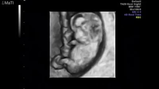
Pregnancy ultrasounds are performed mainly using transabdominal ultrasound. For many women, especially after 8 weeks gestation, sufficient information about the baby may be obtained with transabdominal ultrasound only. However, in the early pregnancy, the developing embryo is very small (at 6 weeks gestation, the baby is only 5-9mm long) and a transvaginal ultrasound may be required to get a better image of the baby. Transvaginal ultrasound is safe and commonly performed during all stages of pregnancy, including the first trimester. It will not harm you or your baby.

New research from Mount Sinai Health System says these surgeries have limited effectiveness and can be economically unjustifiable when they're done on patients with less severe symptoms.
» Subscribe to NBC News: http://nbcnews.to/SubscribeToNBC
» Watch more NBC video: http://bit.ly/MoreNBCNews
NBC News is a leading source of global news and information. Here you will find clips from NBC Nightly News, Meet The Press, and original digital videos. Subscribe to our channel for news stories, technology, politics, health, entertainment, science, business, and exclusive NBC investigations.
Connect with NBC News Online!
Visit NBCNews.Com: http://nbcnews.to/ReadNBC
Find NBC News on Facebook: http://nbcnews.to/LikeNBC
Follow NBC News on Twitter: http://nbcnews.to/FollowNBC
Follow NBC News on Google+: http://nbcnews.to/PlusNBC
Follow NBC News on Instagram: http://nbcnews.to/InstaNBC
Follow NBC News on Pinterest: http://nbcnews.to/PinNBC
New Study Questions Effectiveness Of Knee Replacement Surgery | NBC Nightly News
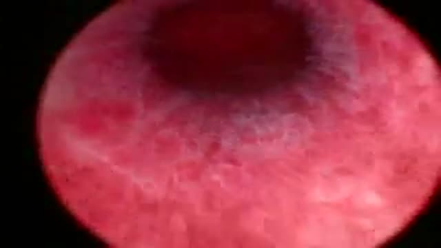
A hystroscopy showing a case of 2 intramural fibroids

The fuel for the process leading to orgasm is testosterone, a hormone produced in steady supply by the testicles. The testicles also make millions of sperm each day, which mature and then are mixed with whitish, protein-rich fluids. These fluids nourish and support the sperm so they can live after ejaculation for a limited time. This mixture of fluid and sperm, known as semen, is what is moved through the urethra and out the penis during orgasm.
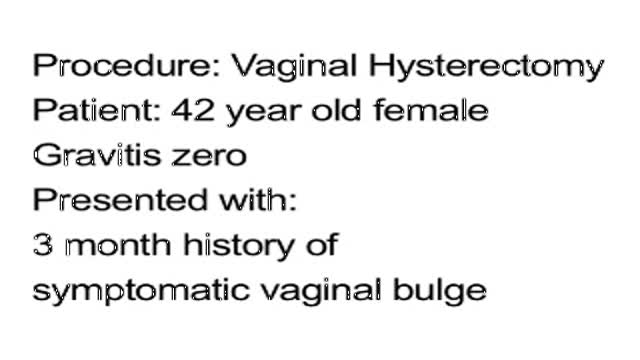
Vaginal Hysterectomy Procedure of a 42 years old female patient with a 3 months history of symptomatic vaginal bulge

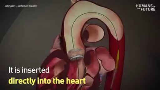
Could this be a viable alternative to open heart surgery?
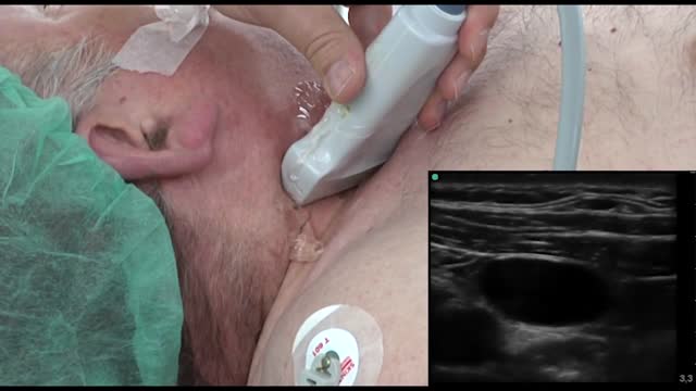
Ultrasound-guided internal jugular cannulation

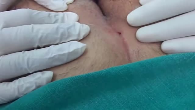
Sports Hernia Self Test (TRY IT)
714-502-4243 | Costa Mesa, CA | http://www.p2sportscare.com
[FREE GIFT] Audio Download
#sportshernia #hernia #hippain
Sports Hernia Diagnosis
What Is A Sports Hernia?
A sports hernia is tearing of the transversalis fascia of the lower abdominal or groin region. A common misconception is that a sports hernia is the same as a traditional hernia. The mechanism of injury is rapid twisting and change of direction within sports, such as football, basketball, soccer and hockey.
The term “sports hernia” is becoming mainstream with more professional athletes being diagnosed. The following are just to name a few:
Torii Hunter
Tom Brady
Ryan Getzlaf
Julio Jones
Jeremy Shockey
If you follow any of these professional athletes, they all seem to have the same thing in common: Lingering groin pain. If you play fantasy sports, this is a major headache since it seems so minor, but it can land a player on Injury Reserve on a moments notice. In real life, it is a very frustrating condition to say the least. It is hard to pin point, goes away with rest and comes back after activity, but is hardly painful enough to make you want to stop. It lingers and is always on your mind. And if you’re looking for my step-by-step sports hernia rehab video course here it is.
One the best definitions of Sport hernias is the following by Harmon:
The phenomena of chronic activity–related groin pain that it is unresponsive to conservative therapy and significantly improves with surgical repair.”
This is truly how sports hernias behave in a clinical setting. It is not uncommon for a sports hernia to be unrecognized for months and even years. Unlike your typical sports injury, most sports medicine offices have only seen a handful of cases. It’s just not on most doctors’ radar. The purpose of this article is not only to bring awareness about sports hernias, but also to educate.
Will you find quick fixes in this article for sports hernia rehab?
Nope. There is no quick fix for this condition, and if someone is trying to sell you one, they are blowing smoke up your you-know-what.
Is there a way to decrease the pain related to sports hernias?
Yes. Proper rehab and avoidance of activity for a certain period of time will assist greatly, but this will not always stop it from coming back. Pain is the first thing to go and last thing to come. Do not be fooled when you become pain-free by resting it. Pain is only one measure of improvement in your rehab. Strength, change of direction, balance and power (just to name a few) are important, since you obviously desire to play your sport again. If you wanted to be a couch potato, you would be feeling better in no time. Watching Sports Center doesn’t require any movement.
Why is this article so long?
There is a lot of information on sports hernias available to you on the web. However, much of the information is spread out all over the internet and hard for athletes to digest due to complicated terminology. This article lays out the foundational terminology you will need to understand what options you have with your injury. We will go over anatomy, biomechanics, rehab, surgery, and even the fun facts. The information I am using is from the last ten years of medical research, up until 2016. We will be making updates overtime when something new is found as well. So link to this page and share with friends. This is the best source for information on sports hernias you will find.
Common Names (or Aliases?) for Sports Hernias
Sportsman’s Hernia
Athletic Pubalgia
Gilmore’s Groin
How Do You Know If You Have A Sports Hernia?
Typical athlete characteristics:
Male, age mid-20s
Common sports: soccer, hockey, tennis, football, field hockey
Motions involved: cutting, pivoting, kicking and sharp turns
Gradual onset
How A Sports Hernia Develops
Chronic groin pain typically happens over time, which is why with sports hernias, we do not hear many stories of feeling a “pop” or a specific moment of injury. It is the result of “overuse” mechanics stemming from a combination of inadequate strength and endurance, lack of dynamic control, movement pattern abnormalities, and discoordination of motion in the groin area.
There is a lot going on in the groin area. There are many muscles, tendons, and fascia pulling in different directions. These contracting structures need to coordinate together for any athletic motion. This perspective is also known as the injury prevention model.

A Lecture Presented by Dr. Mostafa Yakoot to Vascular Surgery Congress. TITLE: SAFETY & EFFICACY OF A NEW HONEY OINTMENT (PEDYPHAR) FOR DIABETIC FOOT ULCERS. Based on the original article in JWC by: Yakoot M, Abdelatif M, Etman M.

AUTO-HEMOTHERAPY IN HERPES CASES. THE STORY OF A DOCTOR IN FERME-NEUVE. CBC NEWS 1977.

Business Insider's Michelle Yan has been nearsighted since she was 9 years old. After laser eye surgery, she has 20/20. She walks us through the pre-surgery steps, the actual surgery, as well as the recovery process.
MORE MEDICAL TECH:
8 Medical Procedures That Are Improving Lives
https://www.youtube.com/watch?v=kTMMrAP6DNI
13 Medical Procedures Changing The Health World
https://www.youtube.com/watch?v=VAR44vnxWis
Lifelike Medical Robot Actually Bleeds
https://www.youtube.com/watch?v=IjnhmcCQLsc
------------------------------------------------------
#Lasik #Surgery #TechInsider
Tech Insider tells you all you need to know about tech: gadgets, how-to's, gaming, science, digital culture, and more.
Visit us at: https://www.businessinsider.com
TI on Facebook: https://www.facebook.com/techinsider
TI on Instagram: https://www.instagram.com/tech_insider/
TI on Twitter: https://twitter.com/techinsider
TI on Amazon Prime: http://read.bi/PrimeVideo
INSIDER on Snapchat: https://insder.co/2KJLtVo
------------------------------------------------------
What It's Like To Get Laser Eye Surgery
A pilonidal sinus (PNS) is a small cyst or abscess that occurs in the cleft at the top of the buttocks. A PNS usually contains hair, dirt, and debris. It can cause severe pain and can often become infected. If it becomes infected, it may ooze pus and blood and have a foul odor. A PNS is a condition that mostly affects men and is also common in young adults. It’s also more common in people who sit a lot, like cab drivers.

External jugular vein cannulation is an integral part of modern medicine and is practiced in virtually every health care setting. Venous access allows sampling of blood, as well as administration of fluids, medications, parenteral nutrition, chemotherapy, and blood products. [1] This topic describes placement of an intravenous (IV) catheter into the external jugular vein. A similar technique can be used for placement of IV catheters at different anatomic sites.

stage of pregnancy 2016
