- Physical Examination
- Surgical Examination
- Ophthalmology
- Clinical Skills
- Orthopedics
- Surgery Videos
- Laparoscopy
- Pediatrics
- Funny Videos
- Cardiothoracic Surgery
- Nursing Videos
- Plastic Surgery
- Otorhinolaryngology
- Histology and Histopathology
- Neurosurgery
- Dermatology
- Pediatric Surgery
- Urology
- Dentistry
- Oncology and Cancers
- Anatomy Videos
- Health and Fitness
- Radiology
- Anaesthesia
- Physical Therapy
- Pharmacology
- Interventional Radiology
- Cardiology
- Endocrinology
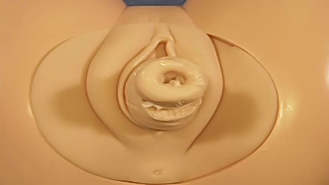
- Gynecology
- Emergency Medicine
- Psychiatry and Psychology
- Childbirth Videos
- General Medical Videos
- Nephrology
- Physiology
- Diet and Food Health
- Diabetes Mellitus
- Neurology
- Women Health
- Osteoporosis
- Gastroenterology
- Pulmonology
- Hematology
- Rheumatology
- Toxicology
- Nuclear Medicine
- Infectious Diseases
- Vascular Disease
- Reproductive Health
- Burns and Wound Healing
- Other
Top videos
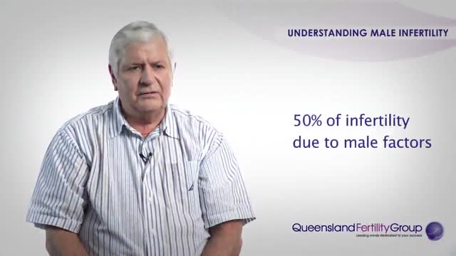
Understanding Male Infertility
A nonsurgical method of treating a ganglion is to drain the fluid from (aspirate) the ganglion sac. Your doctor can do this in the office using the following procedure: The ganglion area is cleaned with an antiseptic solution. A local anesthetic is injected into the ganglion area to numb the area. When the area is numb, the ganglion sac is punctured with a sterile needle. The fluid is drawn out of the ganglion sac. The ganglion collapses. A bandage and, in some cases, a splint are used for a few days to limit movement and prevent the ganglion sac from filling again. Treating a ganglion by draining the fluid with a needle may not work because the ganglion sac remains intact and can fill again, causing the ganglion to return. For this reason, your doctor may puncture the sac with the needle 3 or 4 times so the sac will collapse completely. Even then, the ganglion is likely to come back.

People with Extremely Large Body Parts
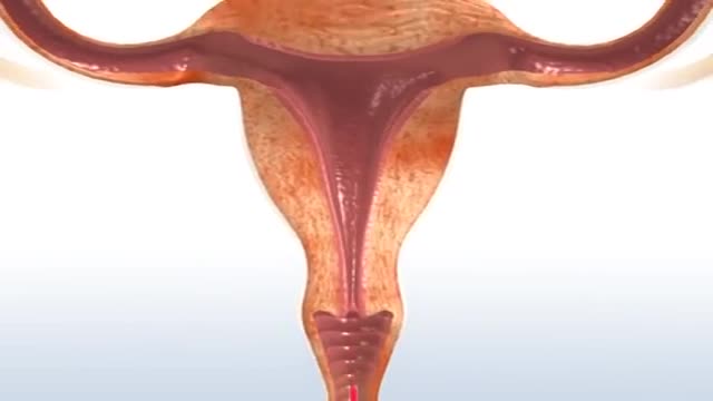
Menstruation is a woman's monthly bleeding. When you menstruate, your body sheds the lining of the uterus (womb). Menstrual blood flows from the uterus through the small opening in the cervix and passes out of the body through the vagina. Most menstrual periods last from 3 to 5 days.

Menorrhagia is the medical term for menstrual periods with abnormally heavy or prolonged bleeding. Although heavy menstrual bleeding is a common concern, most women don't experience blood loss severe enough to be defined as menorrhagia. With menorrhagia, you can't maintain your usual activities when you have your period because you have so much blood loss and cramping. If you dread your period because you have such heavy menstrual bleeding, talk with your doctor. There are many effective treatments for menorrhagia.
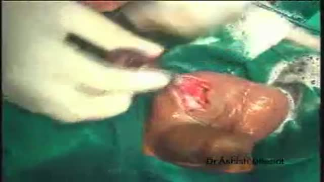
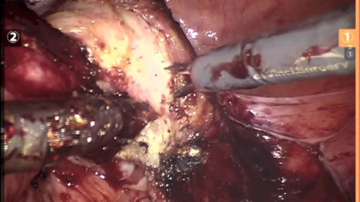
Orchidectomy and Orchidopexy in Testicular Torsion
Cerclage is indicated in a patient with a history of painless cervical dilation and a second trimester loss. It is also indicated in a patient with a history of preterm birth and a short cervix found on ultrasound between 16-24 weeks gestation. Cerclage placement occurs after the first trimester in case the pregnancy is genetically abnormal and would likely result in a first trimester loss.
Hysterectomy is the surgical removal of the uterus. It ends menstruation and the ability to become pregnant. Depending on the reason for the surgery, a hysterectomy may also involve the removal of other organs and tissues such as the ovaries and/or fallopian tubes.
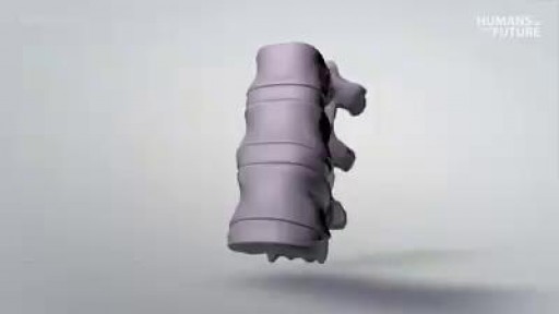
3D printing a titanium vertebrae
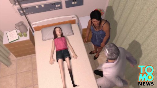
Watch that video of a Snake bite causes girl’s leg to rot away with necrosis
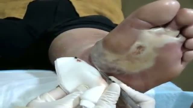
See http://nursing-resource.com for more on debridement.
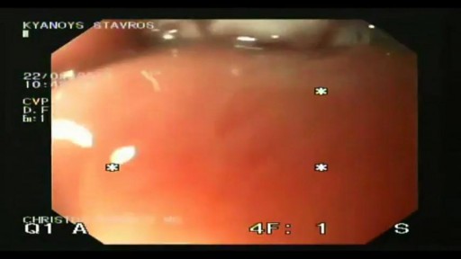
Barrett's esophagus is a complication of chronic (long lasting) and usually severe gastrointestinal reflux disease (GERD), but occurs in only a small percentage of patients with GERD. Criteria are needed for screening patients with GERD for Barrett's esophagus. Until validated criteria are available, it seems reasonable to do screening endoscopies in GERD patients who cannot be taken off acid suppression therapy after two to three years. The diagnosis of Barrett's esophagus rests upon seeing (at endoscopy) a pink esophageal lining that extends a short distance (usually less than 2.5 inches) up the esophagus from the gastroesophageal junction and finding intestinal type cells (goblet cells) on biopsy of the lining. There is a small but definite increased risk of cancer of the esophagus (adenocarcinoma) in patients with Barrett's esophagus.
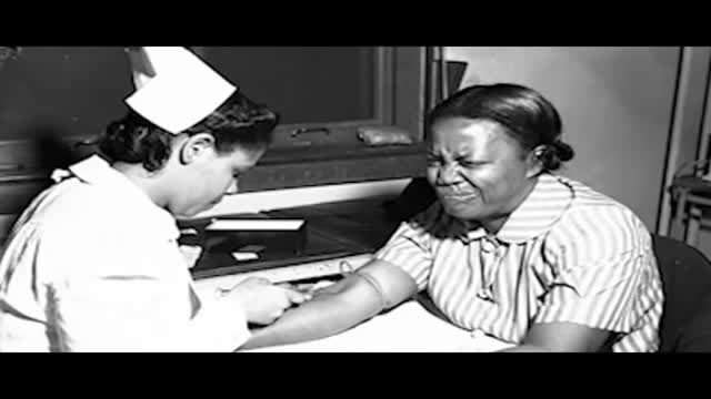
If it gets more severe and causes symptoms, your low hemoglobin count may indicate you have anemia. A low hemoglobin count is generally defined as less than 13.5 grams of hemoglobin per deciliter (135 grams per liter) of blood for men and less than 12 grams per deciliter (120 grams per liter) for women.
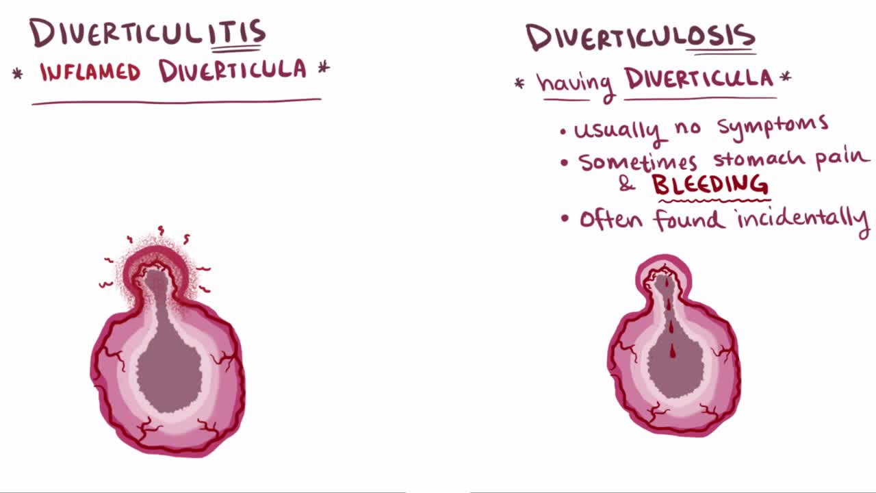
What are diverticula? Diverticula are outpouchings that most commonly happen in the sigmoid colon of the large intestine. The presence of a diverticulum is defined as diverticulosis, whereas diverticulitis describes an inflamed diverticulum
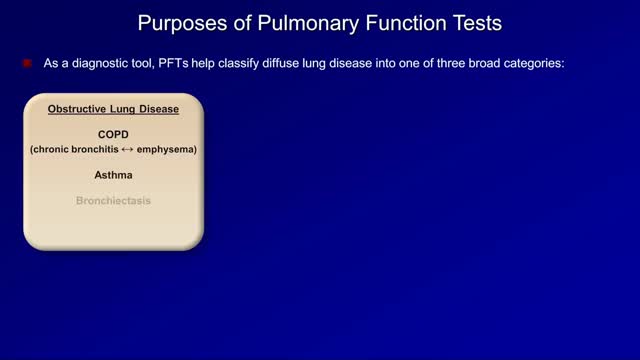
Pulmonary function tests are a broad range of tests that measure how well the lungs take in and exhale air and how efficiently they transfer oxygen into the blood. Spirometry measures how well the lungs exhale.
Swelling is a typical symptom of lymphedema and commonly affects legs and arms. Compression stockings work to encourage the movement of lymph out of an affected limb. Lymphedema is incurable. However, treatment can help reduce the swelling and pain
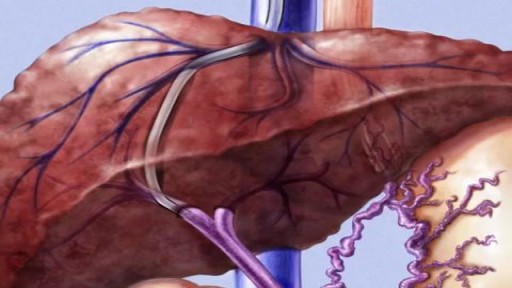
ransjugular intrahepatic portosystemic shunt (TIPS) is a procedure to create new connections between two blood vessels in your liver. You may need this procedure if you have severe liver problems.
Chronic Myeloid Leukemia Diagnosis and Treatment
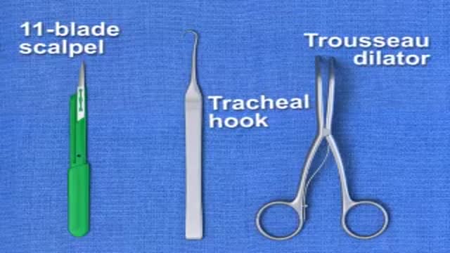
Brief animation demonstrating emergency surgical cricothyrotomy; created with Lightwave 9.3
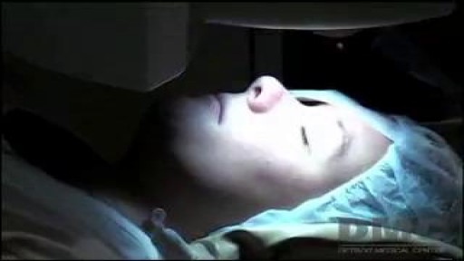
Lasik eye surgery at the Detroit Medical Center's Advanced Laser and Clear Vision Center offer patients pain-free, life-changing procedures that correct nearsightedness, farsightedness, and astigmatism. ~ Detroit Medical Center
