Top videos
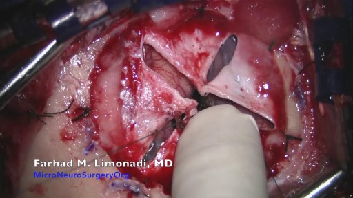
Glioblastoma is a type of astrocytoma, a cancer that forms from star-shaped cells in the brain called astrocytes. In adults, this cancer usually starts in the cerebrum, the largest part of your brain
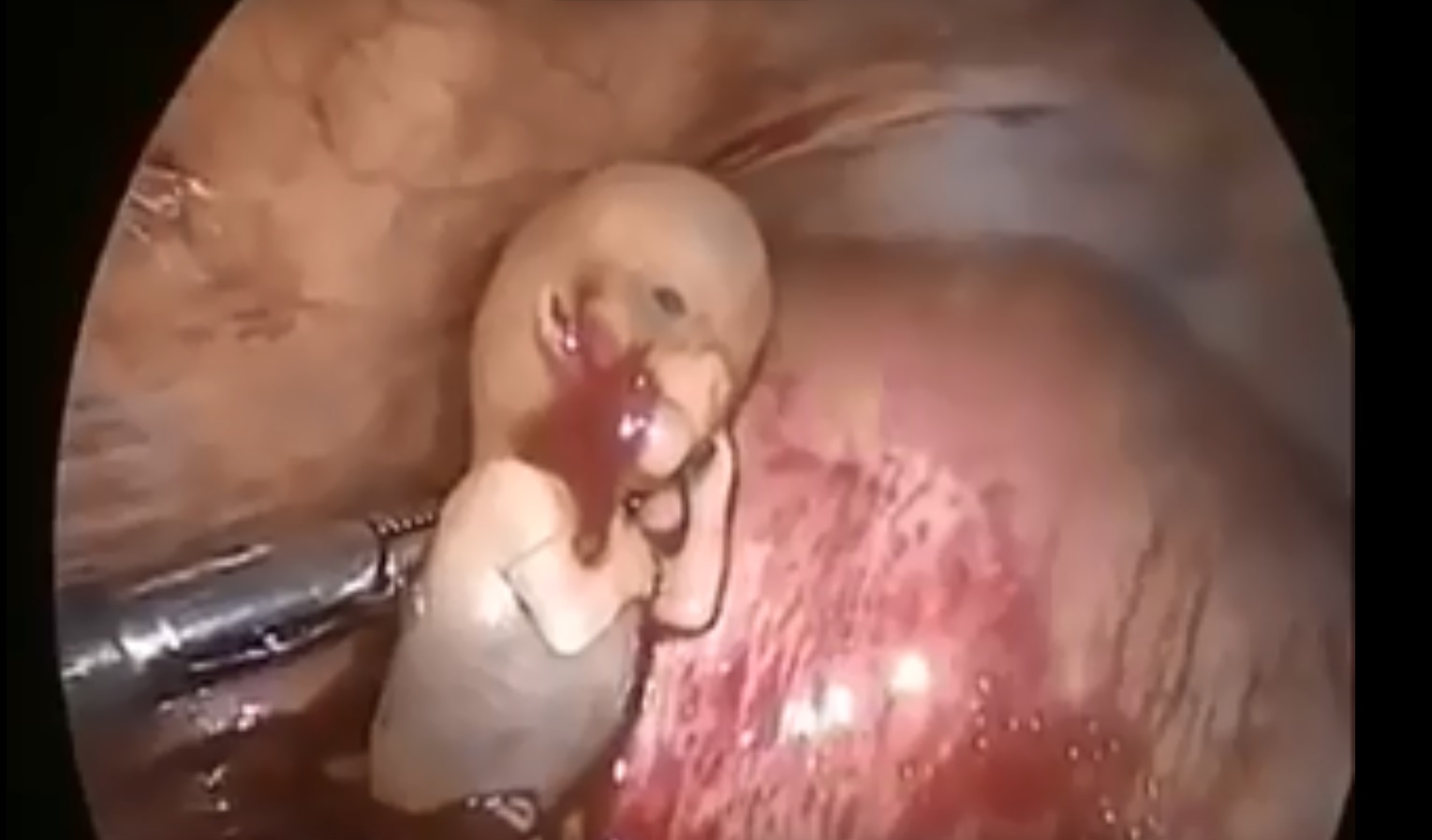
Watch that Ectopic Pregnancy Abortion Surgery
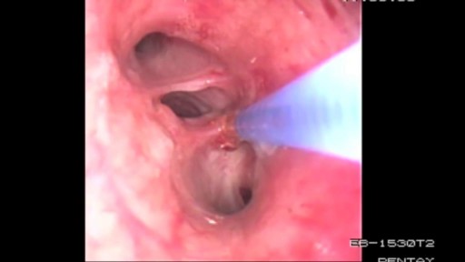
Bronchoscopy Procedure - See inside the lungs!
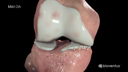
Cartilage is a slippery tissue that provides a smooth surface for joint motion and acts as a cushion between the bones. Synovium is soft, and it lines the joints. It produces fluid, called synovial fluid, for lubrication, and it supplies nutrients and oxygen to the cartilage. As these functions break down, they no longer protect the bones of the knee joint, and bone damage occurs. OA of the knee can cause pain and stiffness. The symptoms worsen over time
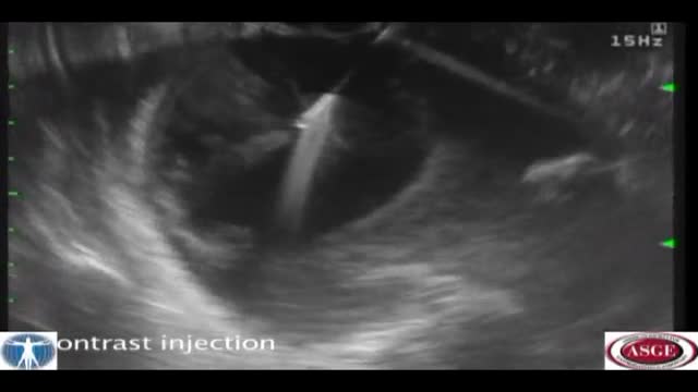
Pancreatic pseudocyst drainage was the first therapeutic application of EUS. The cyst is punctured under ultrasound guidance, contrast injected, and a guidewire inserted. Initial dilation to 8mm is performed over the wire The EUS scope is then exchanged over the wire for a forward viewing endoscope.... A second dilation to 18mm is performed. This enables entry of the endoscope into the cyst perform cystoscopy, debridement if necessary, and insertion of multiple large bore double pigtail stents. The curved linear array-or CLA—echoendoscope has oblique viewing optics located proximal to an oblique scanning transducer. The accessory exits from the shaft of the echoendoscope at an ablique angle, adjustable between 15 and 30 degrees. There are several technical limitations using this echoendoscope. The oblique angle of exit results in a weekend transfer of force when advancing the accessory, difficult deployment of larger bore accessories, and in instrument tunneling effect relative to the bowel wall. There is the potential loss of access during endoscope exchange. A novel CLA echoendoscope was developed by the Olympus Corporation that shifts the orientation of endoscopic and ultrasound views from oblique to forward viewing. The channel is therapeutic at 3.7mm Note that the working channel is located adjacent to the ultrasound transducer at the endoscope tip. The accessory exits the working channel in the axis of the shaft. Shown here are balloon inflation and deployment of a Dormia basket. We report on the use of the prototype forward viewing echoendoscope in six consecutive patients who were referred for pancreatic cyst drainage. Here you see endoscopic view-indistinguisable from that of a gastroscope-showing a bulge where the cyst impinges against the posterior gastric wall. Power Doppler is switched on and highlights multiple vessels interposed in the wall This allows selection of a safe vessel-free window for a cyst puncture A 19 G needle is advanced into the cyst lumen. A sample of contents is aspirated for fluid analysis. A guidewire under ultrasound guidance into the cyst. An 18mm balloon is coaxially thread over the wire and advanced across the cyst wall, Note that resistance is encountered, but the forward transfer of force overcome this. The dilation is performed under forward viewing endoscopuc and ultrasound guidance. As the balloon is maximally inflated we see the cystgastrostomy open up. The balloon is then deflated while simultaneously advancing the scope into the cyst cavity. Cystoscopy isnow performed showing the cyst contents to be filled with pasty wall-adherent necroses. Pulsed power Doppler is switched on we can see and hear arterial flow vessels within the wall of the cyst. This identifies sensitive areas at bleeding risk when performing debridement In this case vigorous water jet irrigation is performed through an accessory water irrigation channel built into the echoendoscope. This issued to clear nonadherent debris. Our experience has shown that it is not necessary to actively remove wall-adherent debris using extraction tools as such Dormia or Roth net basket to achieve cyst resolution. Three large bore 10 Fr double pigtail stents are now inserted into the cyst under direct endoscopic guidance. The first stent is delivered over a guide catheter. The second stent. And the third stent All three stents are deployed. Finally, a nasocystic catheter is inserted for maintenance irrigation. In another patient we used the Cook Cystome to perform cystgastrostomy. We have found the Cystotome easy to delivery through the forward viewing echoendoscope. As shown, we advance the Cystotome into the cyst while applying diathermy. This is performed under and endoscopic guidance, entering the cyst at a near perpendicular orientation. After entry, the Cystotome is removed and cyst fluid gushes from the cystagastrotomy site.
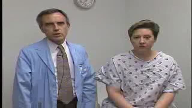
Breast Exam Demonstration
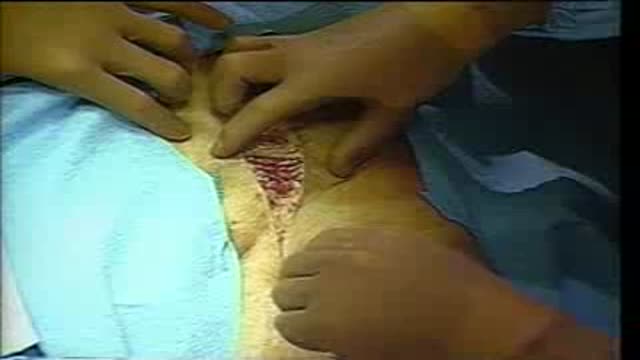
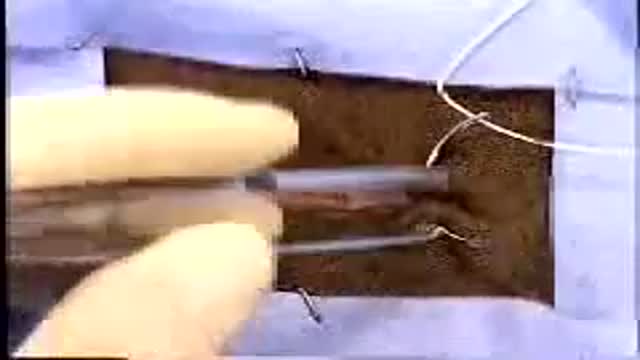
A surgical video showing Femoro-Popliteal Bypass with a Saphenous Vein Graft
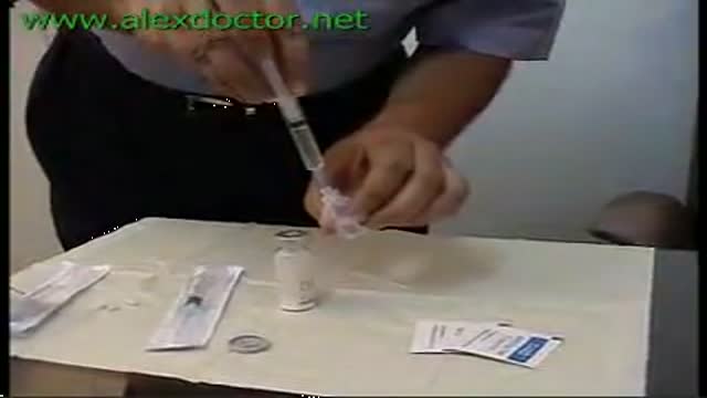
Preparing the Syringe for Injection

Watch that Female Foley Catheter Insertion Procedure
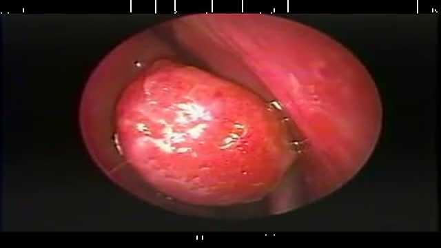
Endoscopic resection of a large right concha bullosa.
Part 2: from Loyola Medical School, Chicago showing clinical examination of the neurological system.
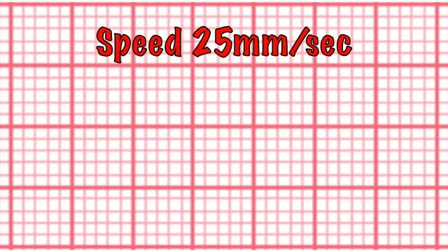
This short course reviews the main features of EKG tracings. A method for analyzing EKGs is also presented. This method includes assessment of rhythm, calculating heart rate, observing P-wave forms, measurement of EKG intervals and segments and the evaluation of other relevant waves.
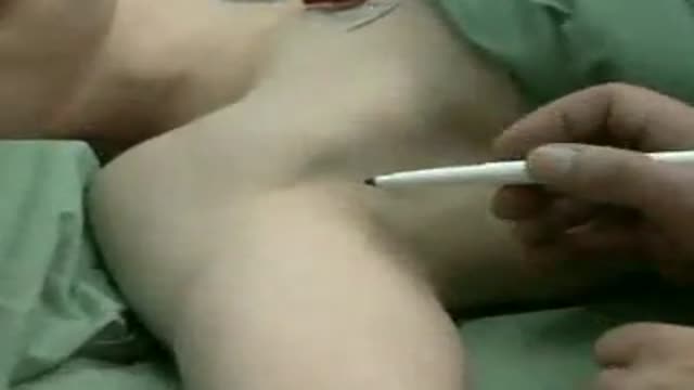
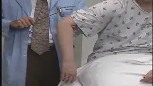
This block is used for procedures of the hand, forearm, and elbow. An injection is given in the patient's axilla (armpit) into a space that surrounds a bundle of nerves that supply feeling to the lower arm. This is usually done with the patient awake with sedation, but can be done with the patient under General Anesthesia.
Possible complications include: Cardiovascular disease. ... Nerve damage (neuropathy). ... Kidney damage (nephropathy). ... Eye damage (retinopathy). ... Foot damage. ... Skin conditions. ... Hearing impairment. ... Alzheimer's disease.

Allergies, what causes them? This animated video reviews the pathophysiology of allergies, what causes them and why the symptoms occur. Food allergies, seasonal allergies and allergies to pollen all occur through a similar mechanism.
Part 5: from Loyola Medical School, Chicago showing clinical examination of the neurological system.

This endoscopy shows a patient with cancer of the larynx, Laryngeal cancer is the most common cancer of the upper respiratory tract. The incidence of laryngeal tumors is closely correlated with smoking, as head and neck tumors occur 6 times more often among cigarette smokers than among nonsmokers. The age-standardized risk of mortality from laryngeal cancer appears to have a linear relationship with increasing cigarette consumption. Death from laryngeal cancer is 20 times more likely for the heaviest smokers than for nonsmokers. It should be suspected in any patient with hoarseness of the voice for three weeks or longer until proven otherwise.
Lembert Pattern Suture

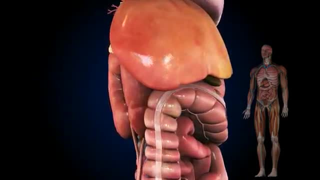
The cat's stomach is a sac-like structure designed to store large volumes of food and continue the digestive process. The esophagus carries food to the stomach, where it enters via a valve-like structure called the cardiac sphincter. On the interior surface of the stomach is a series of folds called gastric folds. These folds function to help grind and digest food. The inner stomach lining secretes acids and enzymes to break down food. Once the initial stomach digestive process is complete, the partially digested food exits the stomach through the pyloric sphincter area and then enters the duodenum (first segment of the small intestine). Once eaten, most food leaves the stomach within twelve hours after entering.
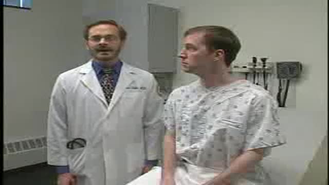
Loyola Respiratory System Exam Part 1 A video from Loyola Medical School, Chicago showing the medical and clinical examination of the respiratory system.
