- Physical Examination
- Surgical Examination
- Ophthalmology
- Clinical Skills
- Orthopedics
- Surgery Videos
- Laparoscopy
- Pediatrics
- Funny Videos
- Cardiothoracic Surgery
- Nursing Videos
- Plastic Surgery
- Otorhinolaryngology
- Histology and Histopathology
- Neurosurgery
- Dermatology
- Pediatric Surgery
- Urology
- Dentistry
- Oncology and Cancers
- Anatomy Videos
- Health and Fitness
- Radiology
- Anaesthesia
- Physical Therapy
- Pharmacology
- Interventional Radiology
- Cardiology
- Endocrinology
- Gynecology
- Emergency Medicine
- Psychiatry and Psychology
- Childbirth Videos
- General Medical Videos
- Nephrology
- Physiology
- Diet and Food Health
- Diabetes Mellitus
- Neurology
- Women Health
- Osteoporosis
- Gastroenterology
- Pulmonology
- Hematology
- Rheumatology
- Toxicology
- Nuclear Medicine
- Infectious Diseases
- Vascular Disease
- Reproductive Health
- Burns and Wound Healing
- Other
Top videos
-Osler-Rendu-Weber syndrome is characterized by multiple telangiectasias and vascular lesions of the CNS.
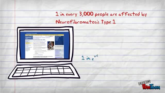
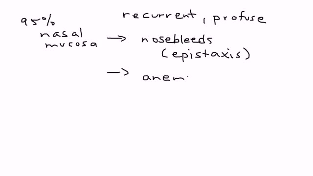
-Hypopigmented spots, in combination with a family history of bilateral deafness, strongly suggest neurofibromatosis type 2 (NF-2), an autosomal-dominant disorder. The spots described actually represent cafe-au-lait spots that are usually hypopigmented (unlike the hyperpigmented cafe-au-lait spots found in NF-1 ). Deafness is caused by bilateral acoustic neuromas, a characteristic neurologic manifestation of the syndrome.
Influenza is a viral infection that attacks your respiratory system — your nose, throat and lungs. Influenza, commonly called the flu, is not the same as stomach "flu" viruses that cause diarrhea and vomiting. For most people, influenza resolves on its own. But sometimes, influenza and its complications can be deadly. People at higher risk of developing flu complications include: Young children under 5, and especially those under 2 years Adults older than 65 Residents of nursing homes and other long-term care facilities Pregnant women and women up to two weeks postpartum People with weakened immune systems People who have chronic illnesses, such as asthma, heart disease, kidney disease and diabetes People who are very obese, with a body mass index (BMI) of 40 or higher Your best defense against influenza is to receive an annual vaccination.

You may initially experience short, mild attacks. But trigeminal neuralgia can progress and cause longer, more-frequent bouts of searing pain. Trigeminal neuralgia affects women more often than men, and it's more likely to occur in people who are older than 50.
How to diagnose digital ulceration in out patient clinic. part II
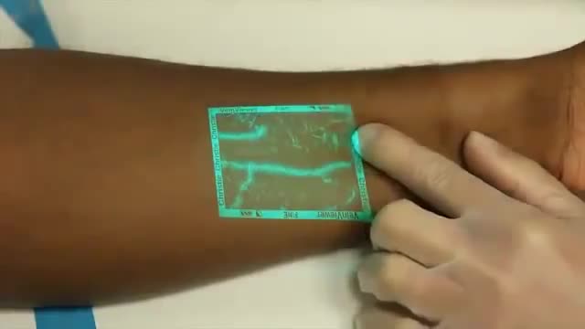
Venipuncture can be a challenging process for medical professionals especially when a patients veins are difficult to see. VeinViewer uses near infrared light to create a digital image of patient vasculature in real time.
Knee pain facts Knee pain is a common problem with many causes, from acute injuries to complications of medical conditions. Knee pain can be localized to a specific area of the knee or be diffuse throughout the knee. Knee pain is often accompanied by physical restriction. A thorough physical examination will usually establish the diagnosis of knee pain. The treatment of knee pain depends on the underlying cause. The prognosis of knee pain is usually good although it might require surgery or other interventions.
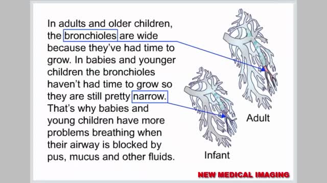
Respiratory syncytial virus (RSV) is a virus that causes infections of the lungs and respiratory tract. It's so common that most children have been infected with the virus by age 2. Respiratory syncytial (sin-SISH-ul) virus can also infect adults. In adults and older, healthy children, the symptoms of respiratory syncytial virus are mild and typically mimic the common cold. Self-care measures are usually all that's needed to relieve any discomfort. Infection with respiratory syncytial virus can be severe in some cases, especially in premature babies and infants with underlying health conditions. RSV can also become serious in older adults, adults with heart and lung diseases, or anyone with a very weak immune system (immunocompromised).
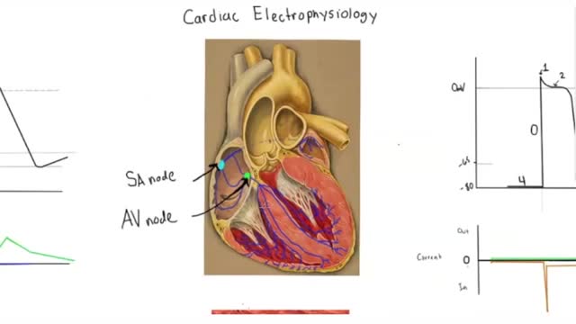
Electrophysiology studies test the electrical activity of your heart to find where an arrhythmia (abnormal heartbeat) is coming from. These results can help you and your doctor decide whether you need medicine, a pacemaker, an implantable cardioverter defibrillator (ICD), cardiac ablation or surgery.
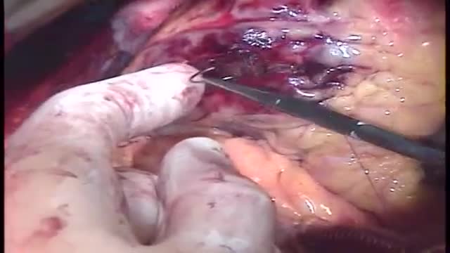
Repair of post-infarction ventricular septal defect (VSD) remains a challenging procedure with a high risk of VSD recurrence. In order to reduce this risk, a double patch and glue technique was introduced in the department in 1986. This surgical technique is hereunder presented. Since 1971, ninety-three patients have been operated on early (≪15 days) after the occurrence of a post-infarction VSD. This retrospective study allows to compare the results of this double patch and glue technique to those obtained with the conventional one, in terms of hospital death and VSD recurrence. The double patch and glue technique avoids recurrence of VSD and plays a part in reducing hospital mortality.
Post-streptococcal GN is a form of glomerulonephritis. It is caused by an infection with a type of streptococcus bacteria. The infection does not occur in the kidneys, but in a different part of the body, such as the skin or throat. The strep bacterial infection causes the tiny blood vessels in the filtering units of the kidneys (glomeruli) to become inflamed. This makes the kidneys less able to filter the urine. Post-streptococcal GN is uncommon today because infections that can lead to the disorder are commonly treated with antibiotics. The disorder may develop 1 to 2 weeks after an untreated throat infection, or 3 to 4 weeks after a skin infection. It may occur in people of any age, but it most often occurs in children ages 6 through 10. Although skin and throat infections are common in children, post-streptococcal GN is a rare complication of these infections. Risk factors include: Strep throat Streptococcal skin infections (such as impetigo)
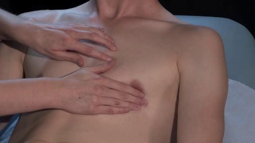
The major elements of the cardiac exam include observation, palpation and, most importantly, auscultation (percussion is omitted). As with all other areas of the physical exam, establishing adequate exposure and a quiet environment are critical. Initially, the patient should rest supine with the upper body elevated 30 to 45 degrees. Most exam tables have an adjustable top. If not, use 2 or 3 pillows. Remember that although assessment of pulse and blood pressure are discussed in the vital signs section they are actually important elements of the cardiac exam.

The window period is the time from infection until a test can detect any change. The average window period with HIV-1 antibody tests is 25 days for subtype B. Antigen testing cuts the window period to approximately 16 days and nucleic acid testing (NAT) further reduces this period to 12 days.[2] Performance of medical tests is often described in terms of: sensitivity: The percentage of the results that will be positive when HIV is present specificity: The percentage of the results that will be negative when HIV is not present. All diagnostic tests have limitations, and sometimes their use may produce erroneous or questionable results. False positive: The test incorrectly indicates that HIV is present in a non-infected person. False negative: The test incorrectly indicates that HIV is absent in an infected person.
Dilated cardiomyopathy is a disease of the heart muscle, usually starting in your heart's main pumping chamber (left ventricle). The ventricle stretches and thins (dilates) and can't pump blood as well as a healthy heart can. The term "cardiomyopathy" is a general term that refers to the abnormality of the heart muscle itself. Dilated cardiomyopathy might not cause symptoms, but for some people it can be life-threatening. A common cause of heart failure — the heart's inability to supply the body with enough blood — dilated cardiomyopathy can also contribute to irregular heartbeats (arrhythmias), blood clots or sudden death. The condition affects people of all ages, including infants and children, but is most common in men ages 20 to 60.
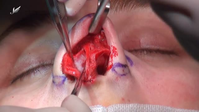
Rhinoplasty enhances facial harmony and the proportions of your nose. It can also correct impaired breathing caused by structural defects in the nose. Rhinoplasty surgery can change: Nose size in relation to facial balance Nose width at the bridge or in the size and position of the nostrils Nose profile with visible humps or depressions on the bridge Nasal tip that is enlarged or bulbous, drooping, upturned or hooked Nostrils that are large, wide, or upturned Nasal asymmetry
Combined Complete Total Gastrectomy with Left Hemipancreatectomy, Splenectomy, Resection of Mesocolon, D3-Lymphadenectomy for Local Advanced Gastric Cancer with Stage IV (T4N2M0).

Menstruation is the time of month when the womb (uterus) sheds its lining and vaginal bleeding occurs. This is known as a menstrual period. Periods vary widely from woman to woman. Some periods are punctual, some are unpredictable. On average, a woman gets her period every 24 to 38 days. A period usually lasts about two to eight days. Irregular periods may require treatment. What Are Irregular Periods? You may have irregular periods if: The time between each period starts to change. You are losing more or less blood during a period than usual. The number of days that your period lasts varies significantly. There are different terms for different types of irregular periods: Irregular Menstrual Bleeding (IrregMB): Bleeding of more than 20 days in individual cycle lengths over a period of one year. Absent Menstrual Bleeding (amenorrhea): No bleeding in a 90-day period. Heavy Menstrual Bleeding (HMB): Excessive menstrual blood loss that interferes with the woman’s physical, emotional, social, and material quality of life and can occur alone or in combination with other symptoms. Heavy and Prolonged Menstrual Bleeding (HPMB): Less common than HMB. It is important to make a distinction from HMB given they may have different etiologies and respond to different therapies. Light Menstrual Bleeding: Based on patient complaint, rarely related to pathology.

https://bit.ly/3HIStRc #shorts
Tracheotomy and tracheostomy are surgical procedures that create an opening in the trachea (windpipe) to help patients breathe when they have difficulty doing so through the nose or mouth. Though they are similar in purpose, there are some key differences between them.
Tracheotomy is a temporary procedure that involves creating a small incision in the trachea to insert a breathing tube. The tube is typically removed once the patient no longer requires it, and the incision heals on its own. Tracheostomy, on the other hand, is a more permanent solution that involves creating a hole in the trachea and inserting a tracheostomy tube, which remains in place for an extended period.
Indications for these procedures include:
Airway obstruction due to trauma, tumors, or infection
Severe respiratory distress or failure
Prolonged mechanical ventilation
Inability to protect the airway due to neurological disorders or impaired consciousness
Steps for performing a tracheotomy and tracheostomy:
Preparation: The patient is positioned, and the neck area is cleaned and draped. Local anesthesia is often administered, although general anesthesia may be used in some cases.
Incision: A small incision is made in the neck, and the muscles and tissues are carefully separated to expose the trachea.
Tracheal opening: A small opening is made in the trachea, typically between the second and third tracheal rings.
Tube insertion: A tracheotomy tube is inserted through the incision and into the trachea for a tracheotomy, while a tracheostomy tube is inserted for a tracheostomy. Both tubes are secured in place.
Confirmation: Proper placement of the tube is confirmed by listening for breath sounds and checking for adequate ventilation.
Pre-operative care typically involves a thorough assessment of the patient's medical history, as well as any necessary imaging studies or lab tests to ensure the procedure is appropriate and safe. Informed consent should be obtained from the patient or their legal representative.
Post-operative care includes monitoring the patient's vital signs, ensuring the tube remains secure and patent, and managing any pain or discomfort. For tracheostomy patients, regular cleaning and maintenance of the stoma (the opening in the trachea) and the tracheostomy tube are essential to prevent infection and other complications. Long-term care may involve speech therapy, respiratory therapy, and support from a multidisciplinary team to address any ongoing needs.
It's crucial to remember that these procedures should only be performed by trained medical professionals in a clinical setting.
for additional information about this procedure check our article @ www.medicalartsshop.com
For more free resources, find us on Pinterest & Facebook pages:
https://www.pinterest.ca/medicalartsofficial/
https://www.facebook.com/Medicalartsofficial
https://www.youtube.com/@medic....alarts?sub_confirmat
https://www.instagram.com/medicalartsofficial/
https://www.tiktok.com/@medicalarts
This video and associated content are for entertainment and educational purposes only!!

Insulin is a hormone made naturally in the pancreas that helps move sugar into the cells of your body. Your cells use the sugar as fuel to make energy. Without enough insulin, sugar stays in your bloodstream, raising your blood sugar. High blood sugar, or hyperglycemia, can lead to the signs and symptoms of diabetes:
Our results in this study of MIPO treated with conventional plates are comparable to the results of the femoral shaft fractures treated with intramedullary nailing. The technique can be used for all femoral shaft fractures. Although the biomechanics of the plate fixation are less stable compared to the intamedullary nail, the mechanical stability is stable enough for bone healing. Healing was rapid, and postoperative care was simplified. The two major complications were malalignment and screw breakage. We recommend using at least three separated screws in each fragment to prevent stress on the screw and screw breakage. Intraoperative limb length, axial alignment, and rotation must be carefully assessed to prevent malalignment. The limitations of our study include lack of a comparison group, retrospective data collection, and no randomisation in outcome evaluation
