- Physical Examination
- Surgical Examination
- Ophthalmology
- Clinical Skills
- Orthopedics
- Surgery Videos
- Laparoscopy
- Pediatrics
- Funny Videos
- Cardiothoracic Surgery
- Nursing Videos
- Plastic Surgery
- Otorhinolaryngology
- Histology and Histopathology
- Neurosurgery
- Dermatology
- Pediatric Surgery
- Urology
- Dentistry
- Oncology and Cancers
- Anatomy Videos
- Health and Fitness
- Radiology
- Anaesthesia
- Physical Therapy
- Pharmacology
- Interventional Radiology
- Cardiology
- Endocrinology
- Gynecology
- Emergency Medicine
- Psychiatry and Psychology
- Childbirth Videos
- General Medical Videos
- Nephrology
- Physiology
- Diet and Food Health
- Diabetes Mellitus
- Neurology
- Women Health
- Osteoporosis
- Gastroenterology
- Pulmonology
- Hematology
- Rheumatology
- Toxicology
- Nuclear Medicine
- Infectious Diseases
- Vascular Disease
- Reproductive Health
- Burns and Wound Healing
- Other
Top videos
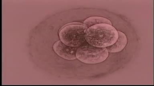
Almost all the cells in your body were produced by mitosis. The only exception is sperm or eggs which are produced by a different type of cell division called meiosis. During fertilization the sperm and egg unite to form a single cell called the zygote which contains chromosomes from both the sperm and egg.
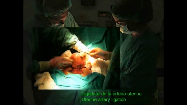
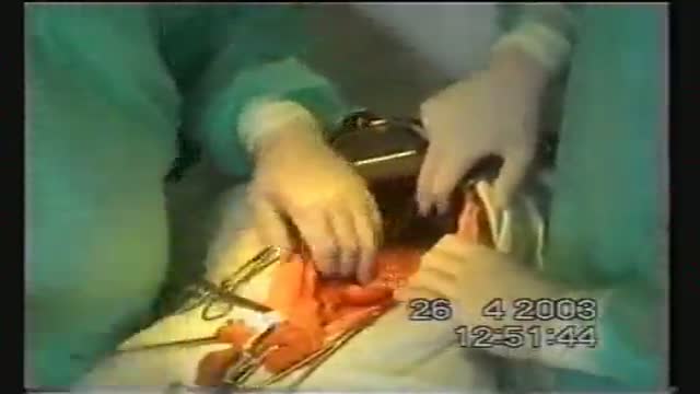
Uterine artery ligation during a C-Section
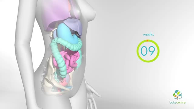
In your first few months of pregnancy, hormones flood your body. Your baby is still tiny but already your body is changing. Your breasts start to swell and may feel tender. Tiredness, nausea and frequent trips to the loo are common pregnancy symptoms.
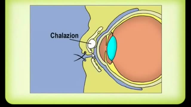
A chalazion is a lump of the lid that is caused by obstruction of the drainage duct of an oil gland within the upper or lower eyelid. This lump may increase in size over days to weeks and may occasionally become red, warm, or painful.
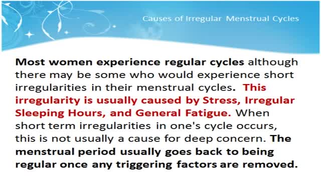
Menstruation is the time of month when the womb (uterus) sheds its lining and vaginal bleeding occurs. This is known as a menstrual period. Periods vary widely from woman to woman. Some periods are punctual, some are unpredictable. On average, a woman gets her period every 24 to 38 days. A period usually lasts about two to eight days. Irregular periods may require treatment. What Are Irregular Periods? You may have irregular periods if: The time between each period starts to change. You are losing more or less blood during a period than usual. The number of days that your period lasts varies significantly. There are different terms for different types of irregular periods: Irregular Menstrual Bleeding (IrregMB): Bleeding of more than 20 days in individual cycle lengths over a period of one year. Absent Menstrual Bleeding (amenorrhea): No bleeding in a 90-day period. Heavy Menstrual Bleeding (HMB): Excessive menstrual blood loss that interferes with the woman’s physical, emotional, social, and material quality of life and can occur alone or in combination with other symptoms. Heavy and Prolonged Menstrual Bleeding (HPMB): Less common than HMB. It is important to make a distinction from HMB given they may have different etiologies and respond to different therapies. Light Menstrual Bleeding: Based on patient complaint, rarely related to pathology.
Occiput or cephalic — the baby's head is down, and the baby is facing the mother's abdomen. This position results in back pain and a prolonged labor. Transverse — the baby is lying crosswise in the uterus, side-to-side over the mother's pelvis, in a horizontal position rather than vertical.
Nasal polyps are associated with inflammation of the lining of your nasal passages and sinuses that lasts more than 12 weeks (chronic rhinosinusitis, also known as chronic sinusitis). However, it's possible — and even somewhat more likely — to have chronic sinusitis without nasal polyps. Nasal polyps themselves are soft and lack sensation, so if they're small you may not be aware you have them. Multiple growths or a large polyp may block your nasal passages and sinuses.

Being diagnosed with ulcerative colitis doesn’t have to stop you from doing the activities you love. Former professional Canadian athletes share their inspiring stories of living well with ulcerative colitis.
Porcelain gallbladder is a condition characterized by calcium salt deposits in the wall of a chronically inflamed gallbladder. The calcifications can be thin or faintly visible, or may be amorphous, patchy, and thick. The gallbladder is generally large, but its size can vary considerably. Most porcelain gallbladders are associated with gallstones. A plain radiograph generally detects these, but computed tomography (CT) has a higher specificity; therefore, a CT scan is performed to confirm the diagnosis. Due to their high risk of gallbladder carcinoma, all patients with porcelain gallbladder should have an elective cholecystectomy.
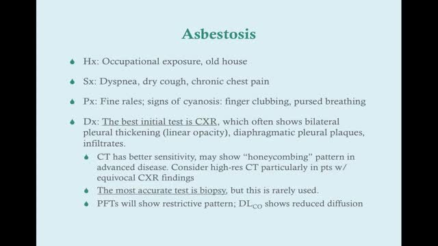
Asbestosis (as-bes-TOE-sis) is a chronic lung disease caused by inhaling asbestos fibers. Prolonged exposure to these fibers can cause lung tissue scarring and shortness of breath. Asbestosis symptoms can range from mild to severe, and usually don't appear until many years after continued exposure. Asbestos is a natural mineral product that's resistant to heat and corrosion. It was used extensively in the past in products such as insulation, cement and some floor tiles. Most people with asbestosis acquired it on the job before the federal government began regulating the use of asbestos and asbestos products in the 1970s. Today, its handling is strictly regulated. Acquiring asbestosis is extremely unlikely if you follow your employer's safety procedures. Treatment focuses on relieving your symptoms.
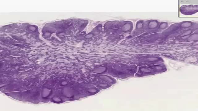
Histology of Lymph Node
Combined Complete Total Gastrectomy with Left Hemipancreatectomy, Splenectomy, Resection of Mesocolon, D3-Lymphadenectomy for Local Advanced Gastric Cancer with Stage IV (T4N2M0).

Full Tummy Tuck 3D Video - http://drlandsman.com
Look great... feel great
•Smart Liposuction + Liposculpture
•Abdominplasty (Tummy Tuck)
+ Full Mini Modified
•Brazilian Lift with Fat Transfer
•Vaginal Aesthetics & Rejuvenation
•Laser Hair Removal
•Full Body Lift
•Thigh lift
•Brachioplasty (Arm Lift) + Short Scar
Expertise in Body Contouring
Board Certified Plastic Surgeon
Expertise in body contouring combines skin excision techniques and advanced fat contouring technology
Weight control personalized training and smoking cessation results in a healthier lifestyle improved shape and longer lasting results
With over 2 decades of experience Dr Lloyd Landsman provides state of the art cosmetic and plastic surgery
Dr Landsman integrates the finest and safest products with the newest procedures
A customized treatment plan is created for each patient utilizing classic surgical and minimally invasive techniques for optimal results
Call for your complimentary consultation to learn how Dr Landsman can help you look your very best
Visit http://drlandsman.com Call 631 864 4111
Main Office 994 W Jericho Tpke Smithtown NY 11787
Affiliates East Islip • Westbury • Jackson Heights • Manhattan
Warning! Do not watch if you are squeamish! SHOW MORE

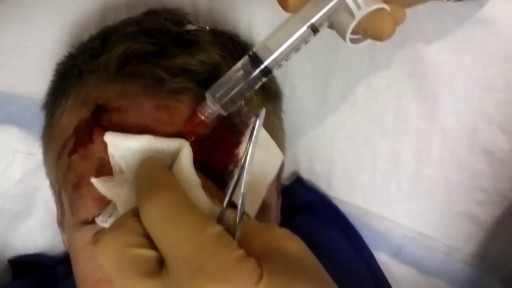
Most minor cuts you can treat yourself -- but know when to see a doctor:
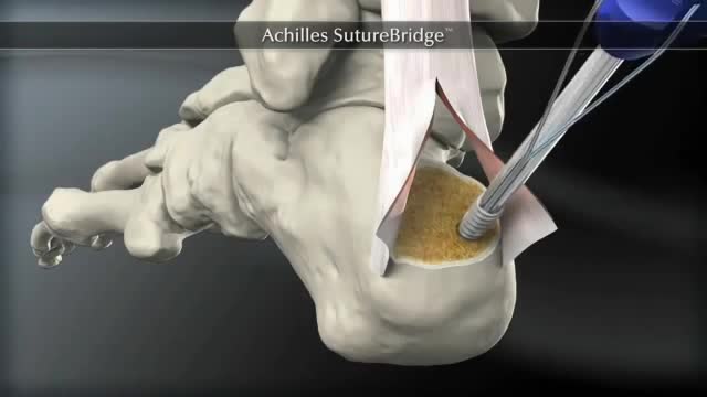
The Arthrex SpeedBridge™ is an innovative soft tissue fixation device used in the treatment of Achilles injuries. While standard anchor fixation of the tendon creates only a single point of compression directly over the anchor, the SpeedBridge enables an hourglass pattern of FiberTape® suture to be laid over the distal end of the tendon. This four-anchor construct enables a true knotless repair and a greater area of compression for the Achilles tendon on the calcaneus, improving stability and possibly allowing for earlier return to normal activities.
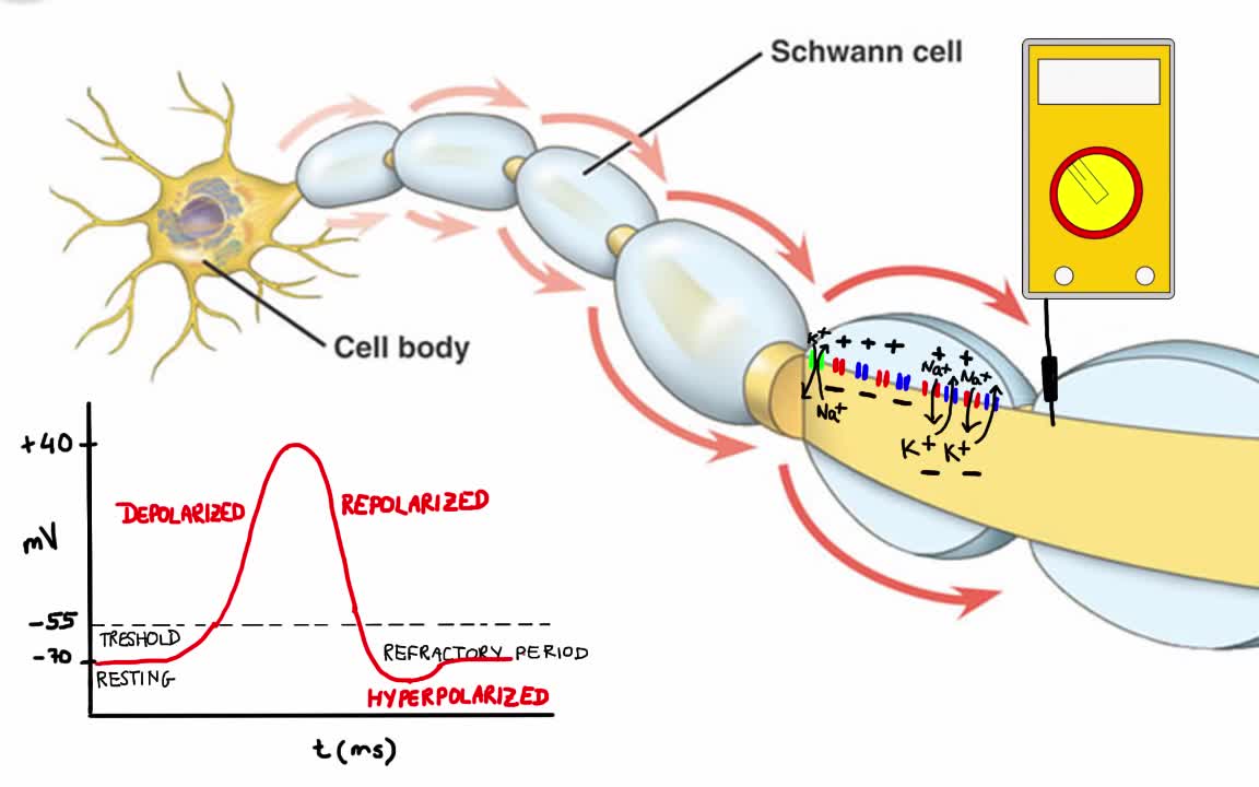
Your body has nerves that connect your brain to the rest of your organs and muscles, just like telephone wires connect homes all around the world. When you want your hand to move, your brain sends signals through your nerves to your hand telling the muscles to contract. But your nerves don’t just say “hand, move.” Instead your nerves send lots of electrical impulses (called action potentials) to different muscles in your hand, allowing you to move your hand with extreme precision.
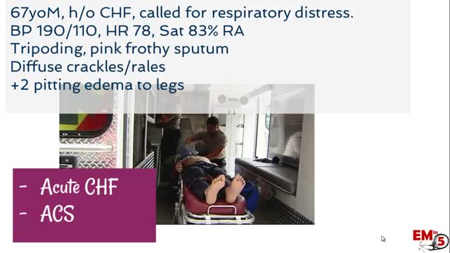
Hypertensive urgency must be distinguished from hypertensive emergency. Urgency is defined as severely elevated blood pressure (ie, systolic >220 mm Hg or diastolic >120 mm Hg) with no evidence of target organ damage.

If you are unhappy with the shape and contours of your breast, then you are not alone. Millions of women around the world are unhappy with their breasts either because they are too small or too big, or too distorted.
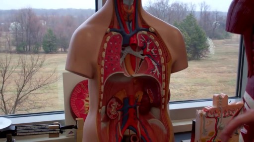
The digestive system is a group of organs working together to convert food into energy and basic nutrients to feed the entire body. Food passes through a long tube inside the body known as the alimentary canal or the gastrointestinal tract (GI tract).
