- Physical Examination
- Surgical Examination
- Ophthalmology
- Clinical Skills
- Orthopedics
- Surgery Videos
- Laparoscopy
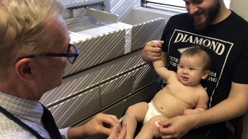
- Pediatrics
- Funny Videos
- Cardiothoracic Surgery
- Nursing Videos
- Plastic Surgery
- Otorhinolaryngology
- Histology and Histopathology
- Neurosurgery
- Dermatology
- Pediatric Surgery
- Urology
- Dentistry
- Oncology and Cancers
- Anatomy Videos
- Health and Fitness
- Radiology
- Anaesthesia
- Physical Therapy
- Pharmacology
- Interventional Radiology
- Cardiology
- Endocrinology
- Gynecology
- Emergency Medicine
- Psychiatry and Psychology
- Childbirth Videos
- General Medical Videos
- Nephrology
- Physiology
- Diet and Food Health
- Diabetes Mellitus
- Neurology
- Women Health
- Osteoporosis
- Gastroenterology
- Pulmonology
- Hematology
- Rheumatology
- Toxicology
- Nuclear Medicine
- Infectious Diseases
- Vascular Disease
- Reproductive Health
- Burns and Wound Healing
- Other
Top videos
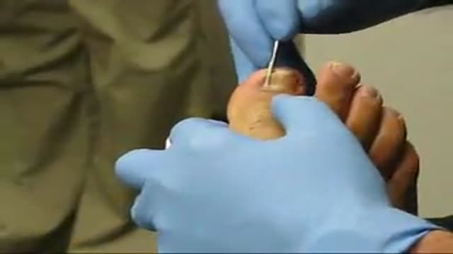
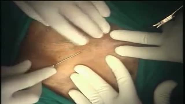
Ingrown Toenail Removal

Treating Hernia with Laparscopic Inguinal Hernia Repair
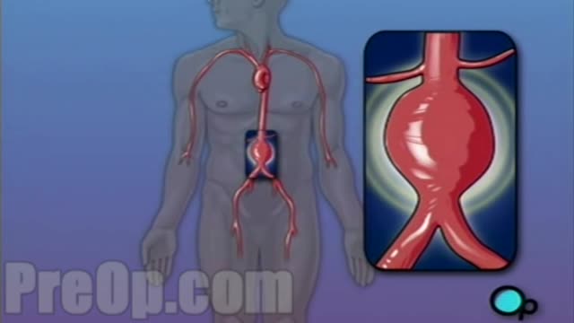
For this surgery, your doctor makes a large incision in the abdomen to expose the aorta. Once he or she has opened the abdomen, a graft can be used to repair the aneurysm. Open repair remains the standard procedure for an abdominal aortic aneurysm repair. Endovascular aneurysm repair (EVAR).
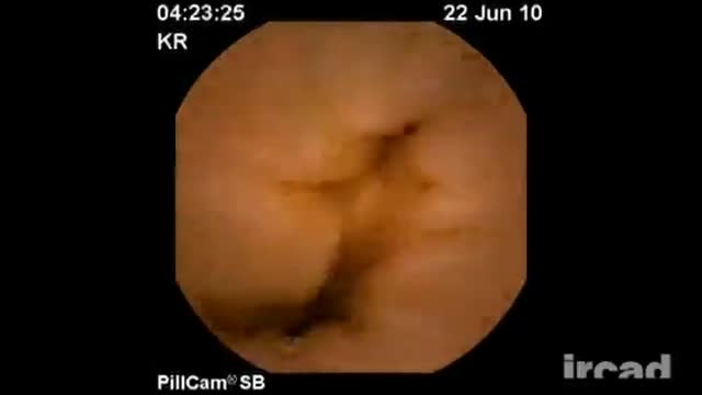
Meckels Diverticulum

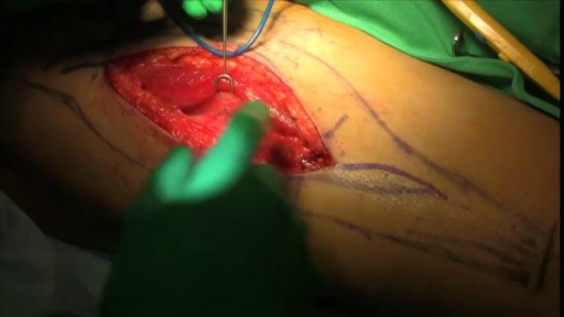
Hip Resurfacing Surgery Videos Welcome to the website of the Asian Regional Center for Hip Resurfacing (ARCH) is a specialized surgical center in Apollo Speciality Hospital Chennai. More than 1350 Hip Resurfacing Surgeries have been performed so far. Asian Regional Center for Hip Resurfacing is the first specialized resurfacing center in Asia. Patients with arthritis and hip pain travel from all over the world travel to ARCH for hip surgery. Hip Resurfacing Surgery has revolutionized hip arthroplasty especially for younger and active patients. Unlike conventional Total Hip Replacement (THR) the hip resurfacing conserves the bone in the hip which would be crucial in younger patients. No restrictions are imposed on the resurfaced hip and the patient can participate in any professional or recreational activity after the surgery.
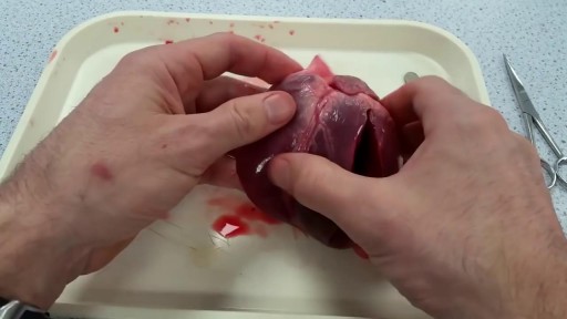
Heart dissection Explaination
Hernia Repair with Prolene Hernia System
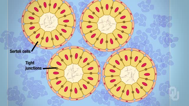
The male reproductive system includes the scrotum, testes, spermatic ducts, sex glands, and penis. These organs work together to produce sperm, the male gamete, and the other components of semen.
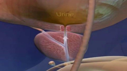
Watch that video of Sperm Formation and Pathway Ejaculation
Infected Hernia Mesh Repair Surgery Video

Traumatic brain injury (TBI) is a nondegenerative, noncongenital insult to the brain from an external mechanical force, possibly leading to permanent or temporary impairment of cognitive, physical, and psychosocial functions, with an associated diminished or altered state of consciousness.
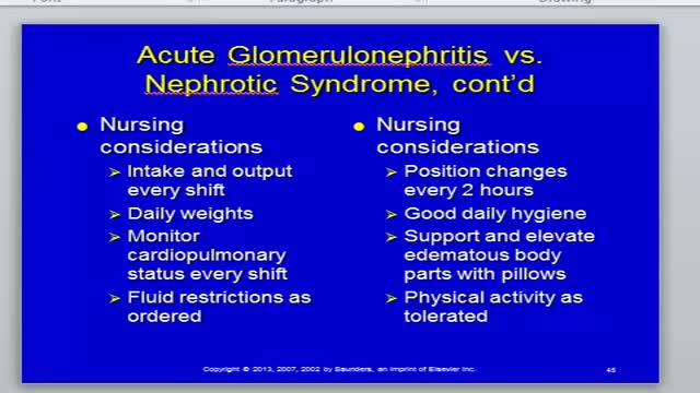
Nephritis and Nephrotic Syndrome

In developing countries, domestic animals (eg, dogs) are common sources of infection. In the United States, bats and wild animals (eg, raccoons) are the most common reservoirs of infection. The acquisition of rabies from bats can occur from an unrecognized bite or a scratch, and possibly by inhalation of aerosolized viral particles. Bats are found in all states except Hawaii, and spelunking (cave exploration) is a risk factor for rabies acquisition from bats.
Back pain during pregnancy is a common complaint — and it's no wonder. You're gaining weight, your center of gravity changes, and your hormones are relaxing the ligaments in the joints of your pelvis. Often, however, you can prevent or ease back pain during pregnancy. Consider seven ways to give pregnancy back pain the boot. 1. Practice good posture As your baby grows, your center of gravity shifts forward. To avoid falling forward, you might compensate by leaning back — which can strain the muscles in your lower back and contribute to back pain during pregnancy. Keep these principles of good posture in mind: Stand up straight and tall. Hold your chest high. Keep your shoulders back and relaxed. Don't lock your knees. When you stand, use a comfortably wide stance for the best support. If you must stand for long periods of time, rest one foot on a low step stool — and take time for frequent breaks. Good posture also means sitting with care. Choose a chair that supports your back, or place a small pillow behind your lower back. 2. Get the right gear Wear low-heeled — not flat — shoes with good arch support. Avoid high heels, which can further shift your balance forward and cause you to fall. You might also consider wearing a maternity support belt. Although research on the effectiveness of maternity support belts is limited, some women find the additional support helpful. 3. Lift properly When lifting a small object, squat down and lift with your legs. Don't bend at the waist or lift with your back. It's also important to know your limits. Ask for help if you need it. 4. Sleep on your side Sleep on your side, not your back. Keep one or both knees bent. Consider using pregnancy or support pillows between your bent knees, under your abdomen and behind your back.
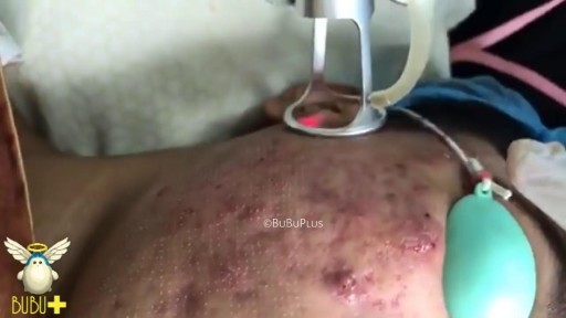
Laser Cystic Acne and Pimples Extraction
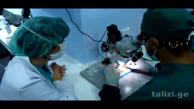
The Talizi Hair Transplantation Clinic offers hair restoration through a painless hair transplantation procedure and guarantees a natural result for an affordable price. 6000 grafts at one session. Hair transplantation surgery combining seamless Follicular Unit Extraction FUE method and Strip Version.

Causes of Polycystic Ovarian Syndrome|| Common gynaecological problems in women Polycystic ovarian syndrome, or PCOS, is a condition where a woman's ovaries and adrenal glands produce more androgens than normal, resulting in increased body hair, acne and irregular periods. While researchers are not certain of the exact cause of PCOS, it is known that an imbalance of the endocrine system is responsible for many of the changes associated with it. However, it is still not known exactly what causes those changes.
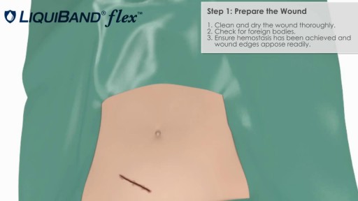
Learn the proper technique for applying LiquiBand Flex Topical Skin Adhesive
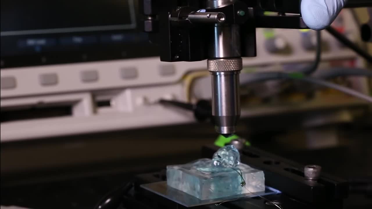
University of California, Berkeley engineers have built the first dust-sized, wireless sensors that can be implanted in the body, bringing closer the day when a Fitbit-like device could monitor internal nerves, muscles or organs in real time.
