- Physical Examination
- Surgical Examination
- Ophthalmology
- Clinical Skills
- Orthopedics
- Surgery Videos
- Laparoscopy
- Pediatrics
- Funny Videos
- Cardiothoracic Surgery
- Nursing Videos
- Plastic Surgery
- Otorhinolaryngology
- Histology and Histopathology
- Neurosurgery
- Dermatology
- Pediatric Surgery
- Urology
- Dentistry
- Oncology and Cancers
- Anatomy Videos
- Health and Fitness
- Radiology
- Anaesthesia
- Physical Therapy
- Pharmacology
- Interventional Radiology
- Cardiology
- Endocrinology
- Gynecology
- Emergency Medicine
- Psychiatry and Psychology
- Childbirth Videos
- General Medical Videos
- Nephrology
- Physiology
- Diet and Food Health
- Diabetes Mellitus
- Neurology
- Women Health
- Osteoporosis
- Gastroenterology
- Pulmonology
- Hematology
- Rheumatology
- Toxicology
- Nuclear Medicine
- Infectious Diseases
- Vascular Disease
- Reproductive Health
- Burns and Wound Healing
- Other
Top videos

Learn about electromagnetic navigation diagnostic bronchoscopy, a new technology used to diagnose small lung cancer tumors as small as a pencil eraser before they have the chance to spread. Cleveland Clinic physician Dr. Thomas Gildea demonstrates how this endobronchial ultrasound procedure, which involves using a small camera probe inserted thru the nose into the lungs, allows doctors to reach possible cancer in the lungs that they could never reliably get to before
Spinal anesthesia is done in a similar way. But the anesthetic medicine is injected using a much smaller needle, directly into the cerebrospinal fluid that surrounds the spinal cord. The area where the needle will be inserted is first numbed with a local anesthetic. Then the needle is guided into the spinal canal, and the anesthetic is injected. This is usually done without the use of a catheter. Spinal anesthesia numbs the body below and sometimes above the site of the injection. The person may not be able to move his or her legs until the anesthetic wears off.
With a portable pump controlled by a wireless handheld device that automatically delivers insulin.
Open Inguinal Hernia Repair Surgery - German Narration

If you’re wondering ‘what’s the cause of my knee pain?’ or ‘what kind of knee pain do I have?’ the position of your knee pain can often tell you what type of knee pain you have.
You confirm this if you know the common symptoms an aggravations for each type of knee problem. So if you want to know ‘why my knee hurts’... here’s a quick look at the most common type of knee problems...
Patellofemoral Pain Syndrome (Or Runner’s Knee) (Old Name: Chondromalacia Patellae)
Infrapatellar Fat Pad Syndrome (Hoffa's Syndrome)
Patella Tendonitis (Jumper’s Knee)
Prepatellar Bursitis
Osgood-Schlatter Disease
Meniscus Tear
Medial Collateral Ligament Tear
Osteoarthritic Knee Pain
Pes Anserine Bursitis.
Iliotibial Band Syndrome
Quadriceps Tendinopathy
Popliteus Strain
Baker’s Cyst
ACL Or PCL Tear/Rupture
---------------------------------------
Check out my channel...
https://youtube.com/@BodyFixExercises
OTHER VIDEOS:
How To Fix Pain In The Front Of The Knee… (Runner's Knee) https://youtu.be/g0qmx_0enAA
Knee Strengthening Exercises To Prevent Knee Pain
https://youtu.be/Pk-ae_lyx7M
How To Treat Patellar Tendinopathy (Jumper’s Knee) & Quadriceps Tendinopathy
https://youtu.be/MkPwsb-rQwU
---------------------------------------
#bodyfixexercises #kneepainrelief #kneepain

A video showing the procedure of Tubular Diskectomy of a herniated disk. Uploaded on MedicalVideos.us.Discussing the management of Sciatica.

Bronchiectasis is an abnormal dilation of the proximal and medium-sized bronchi (>2 mm in diameter) caused by weakening or destruction of the muscular and elastic components of the bronchial walls. Affected areas may show a variety of changes, including transmural inflammation, edema, scarring, and ulceration, among other findings. Distal lung parenchyma may also be damaged secondary to persistent microbial infection and frequent postobstructive pneumonia. Bronchiectasis can be congenital but is most often acquired.[9] Congenital bronchiectasis usually affects infants and children. These cases result from developmental arrest of the bronchial tree. Acquired forms occur in adults and older children and require an infectious insult, impairment of drainage, airway obstruction, and/or a defect in host defense. The tissue is also damaged in part by the host response of neutrophilic proteases, inflammatory cytokines, nitric oxide, and oxygen radicals. This results in damage to the muscular and elastic components of the bronchial wall. Additionally, peribronchial alveolar tissue may be damaged, resulting in diffuse peribronchial fibrosis.[12] The result is abnormal bronchial dilatation with bronchial wall destruction and transmural inflammation. The most important functional finding of altered airway anatomy is severely impaired clearance of secretions from the bronchial tree. Impaired clearance of secretions causes colonization and infection with pathogenic organisms, contributing to the purulent expectoration commonly observed in patients with bronchiectasis. The result is further bronchial damage and a vicious cycle of bronchial damage, bronchial dilation, impaired clearance of secretions, recurrent infection, and more bronchial damage
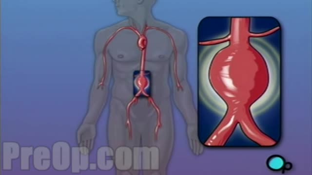
For this surgery, your doctor makes a large incision in the abdomen to expose the aorta. Once he or she has opened the abdomen, a graft can be used to repair the aneurysm. Open repair remains the standard procedure for an abdominal aortic aneurysm repair. Endovascular aneurysm repair (EVAR).
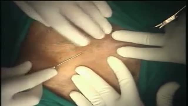
Hernia Repair with Prolene Hernia System

Most healthy children are inattentive, hyperactive or impulsive at one time or another. It’s normal for preschoolers to have short attention spans and be unable to stick with one activity for long. Even in older children and teenagers, attention span often depends on the level of interest. The same is true of hyperactivity. Young children are naturally energetic — they often are still full of energy long after they’ve worn their parents out. In addition, some children just naturally have a higher activity level than others do. Children should never be classified as having ADHD just because they’re different from their friends or siblings. Children who have problems in school but get along well at home or with friends are likely struggling with something other than ADHD. The same is true of children who are hyperactive or inattentive at home, but whose schoolwork and friendships remain unaffected.
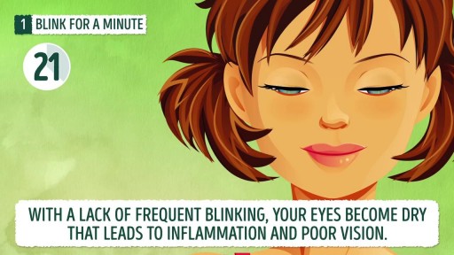
How to improve your eyesight at home? Exercising your eyes is one of those simple things that very few people do. However, it can help you maintain excellent vision. Here are 10 exercises that will take you no more than ten minutes to do. You can give them a try right now while watching this video – we are going to do all of them with you! Exercise #1. Blink for a minute. Exercise #2. Rotate your head while staring ahead. Exercise #3. Look to your right and left. Exercise #4. Close your eyes and relax. Exercise #5. Move your gaze in different directions. Exercise #6. Close and open your eyes. Exercise #7. Push against your temples with your fingers. Exercise #8. Draw geometric figures with your gaze. Exercise #9. Move your eyeballs up and down. Exercise #10. Strengthen your eyes’ near and far focusing.
Meckels Diverticulum

In a normal hip, the ball at the upper end of the thighbone (femur) fits firmly into the socket, which is part of the large pelvis bone. In babies and children with developmental dysplasia (dislocation) of the hip (DDH), the hip joint has not formed normally.
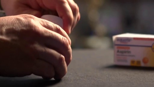
You should not use aspirin if you have a bleeding disorder such as hemophilia, a recent history of stomach or intestinal bleeding, or if you are allergic to an NSAID (non-steroidal anti-inflammatory drug) such as Advil, Motrin, Aleve, Orudis, Indocin, Lodine, Voltaren, Toradol, Mobic, Relafen, Feldene, and others. Do not give this medication to a child or teenager with a fever, flu symptoms, or chicken pox. Salicylates can cause Reye's syndrome, a serious and sometimes fatal condition in children.
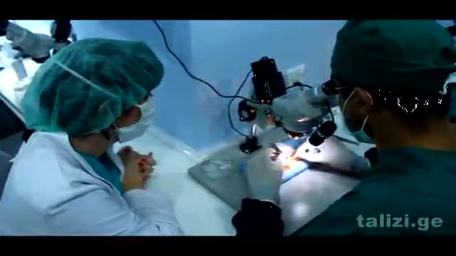
The Talizi Hair Transplantation Clinic offers hair restoration through a painless hair transplantation procedure and guarantees a natural result for an affordable price. 6000 grafts at one session. Hair transplantation surgery combining seamless Follicular Unit Extraction FUE method and Strip Version.
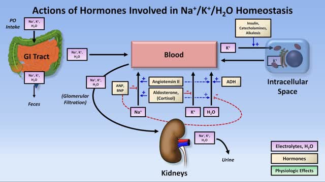
Low potassium (hypokalemia) refers to a lower than normal potassium level in your bloodstream. Potassium is a chemical (electrolyte) that is critical to the proper functioning of nerve and muscles cells, particularly heart muscle cells. Normally, your blood potassium level is 3.6 to 5.2 millimoles per liter (mmol/L). A very low potassium level (less than 2.5 mmol/L) can be life-threatening and requires urgent medical attention.

Traumatic penile injury can be due to multiple factors. Penile fracture, penile amputation, penetrating penile injuries, and penile soft tissue injuries are considered urologic emergencies and typically require surgical intervention. The goals of treatment for penile trauma are universal: preservation of penile length, erectile function, and maintenance of the ability to void while standing. Traumatic injury to the penis may concomitantly involve the urethra.[1, 2] Urethral injury and repair is beyond the scope of this article but details can be found in Urethral Trauma. Penile fracture Penile fracture is the traumatic rupture of the corpus cavernosum. Traumatic rupture of the penis is relatively uncommon and is considered a urologic emergency.[3] Sudden blunt trauma or abrupt lateral bending of the penis in an erect state can break the markedly thinned and stiff tunica albuginea, resulting in a fractured penis. One or both corpora may be involved, and concomitant injury to the penile urethra may occur. Urethral trauma is more common when both corpora cavernosa are injured.[4] Penile rupture can usually be diagnosed based solely on history and physical examination findings; however, in equivocal cases, diagnostic cavernosography or MRI should be performed. Concomitant urethral injury must be considered; therefore, preoperative retrograde urethrographic studies should generally be performed. See the images below.

Mitosis is the process in which a eukaryotic cell nucleus splits in two, followed by division of the parent cell into two daughter cells. The word "mitosis" means "threads," and it refers to the threadlike appearance of chromosomes as the cell prepares to divide.
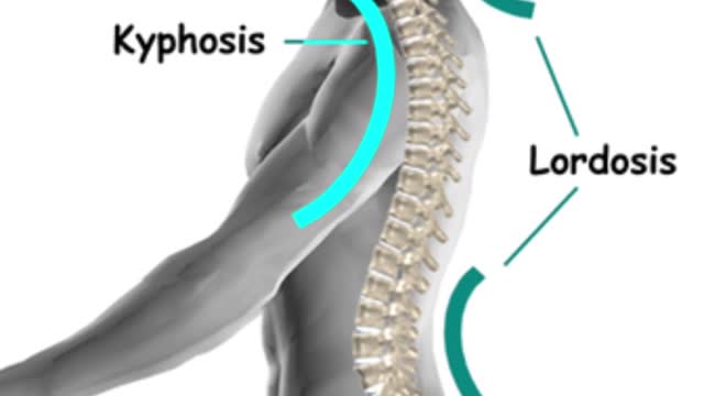
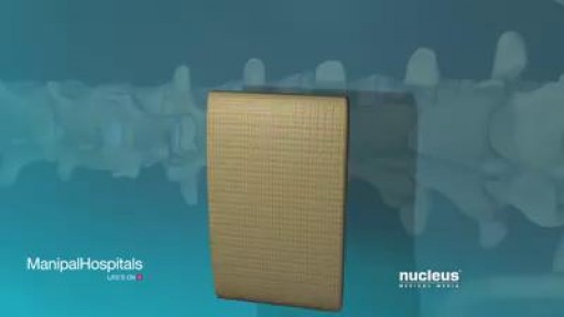
The gradual curves of the human spine allow the body to absorb many shocks and stresses in daily life. It’s a delicate balance, though, and if part of the spine curves too much, pain and limited mobility may result.
Watch that video of Penile Lengthening and Girth Enhancement Plastic Surgery
