- Physical Examination
- Surgical Examination
- Ophthalmology
- Clinical Skills
- Orthopedics
- Surgery Videos
- Laparoscopy
- Pediatrics
- Funny Videos
- Cardiothoracic Surgery
- Nursing Videos
- Plastic Surgery
- Otorhinolaryngology
- Histology and Histopathology
- Neurosurgery
- Dermatology
- Pediatric Surgery
- Urology
- Dentistry
- Oncology and Cancers
- Anatomy Videos
- Health and Fitness
- Radiology
- Anaesthesia
- Physical Therapy
- Pharmacology
- Interventional Radiology
- Cardiology
- Endocrinology
- Gynecology
- Emergency Medicine
- Psychiatry and Psychology
- Childbirth Videos
- General Medical Videos
- Nephrology
- Physiology
- Diet and Food Health
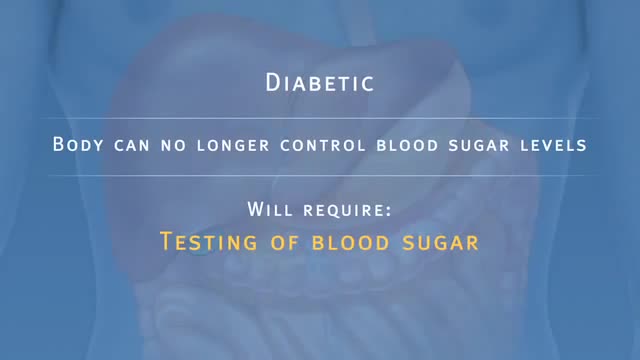
- Diabetes Mellitus
- Neurology
- Women Health
- Osteoporosis
- Gastroenterology
- Pulmonology
- Hematology
- Rheumatology
- Toxicology
- Nuclear Medicine
- Infectious Diseases
- Vascular Disease
- Reproductive Health
- Burns and Wound Healing
- Other
Top videos
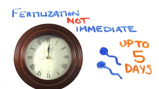
Emergency contraception is a method of birth control you can use if you had sex without using birth control or if your birth control method did not work correctly. You must use emergency contraception as soon as possible after unprotected sex. Emergency contraception pills are different from the abortion pill. If you are already pregnant, emergency contraception pills do not stop or harm your pregnancy. Emergency contraception has also been called the "morning-after pill," but you do not need to wait until the morning after unprotected sex to take it. Emergency contraception is not meant to be used for regular birth control. Talk to your doctor or nurse about regular birth control to help prevent pregnancy. Nearly half of all pregnancies in the United States are unplanned.1
Ever heard medical terms like MRI or EKG? Funny speaker for nurses and doctors and all-around healthcare speaker Dr. Brad Nieder discusses the funny medical jargon he's encountered during his medical career.
He jokes about medical acronyms and big healthcare terms. His funny medical humor makes the conference attendees burst with laughter and he reads the medical definition for "laugh."
As an experienced physician and keynote speaker, he's perfect for any in-person or virtual conference or event. He's also a great healthcare speaker to bring in for continuing medical education (cme) units!
Learn more about Brad's keynote and virtual speaking, and book him for your next conference or virtual event: https://www.HealthyHumorist.com
Find Dr. Brad on social media:
https://www.facebook.com/HealthyHumor...
https://www.linkedin.com/in/BradNieder
https://twitter.com/HealthyHumorist
https://www.youtube.com/c/BradNiederMD
https://vimeo.com/BradNieder
Brad Nieder, MD, CSP*
The Healthy Humorist
Doctor, Keynote Speaker, Clean Comedian
*CSP=Certified Speaking Professional
"Medical Lingo"
From the DVD "The Healthy Humorist in Orlando: Laughter is the Best Medicine"
Animation explaining the pancreatic auto islet transplantation process with complete removal of the pancreas to treat pancreatitis.

Watch that video of a Terrible Bodybuilder's Colon Contains 10 lbs of Meat Worms
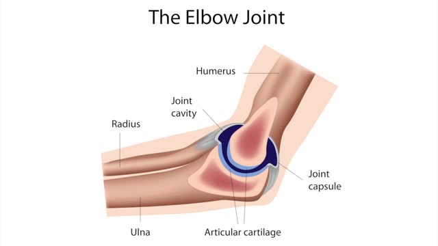
Golfer's elbow causes pain that starts on the inside bump of the elbow, the medial epicondyle. Wrist flexors are the muscles of the forearm that pull the hand forward. The wrist flexors are on the palm side of the forearm. Most of the wrist flexors attach to one main tendon on the medial epicondyle.
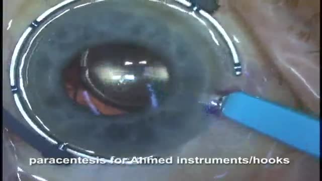
Here Drs Oetting and Shriver of the University of Iowa demonstrate the McCannel technique of fixing an IOL to the iris. In this video both the standard McCannel suture retrieval technique and the Siepser/Chang modifed technique are demonstrated. A 10-O prolene with a long curved ctc-6 needle is u...sed to place a suture through the iris and under an 3 piece IOL haptic. Using the standard technique the two ends of the suture are retrieved through a common paracentesis near the fixation site and tied externally. The other haptic is tied using the Siepser sliding knot technique as described by Chang for this indication with an internal knot. The standard technique is a bit easier but does not allow as thight a knot for fixation of the iris to the haptic.
Watch that video to learn everything about Hemorrhoids Repairing Surgery
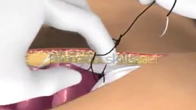
Laparotomy Closure Abdomen Animation
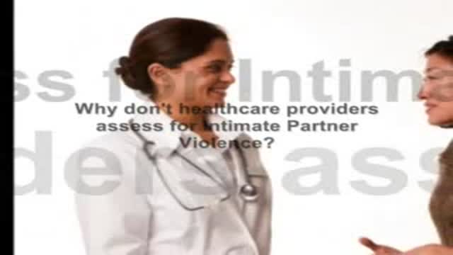
Healthcare providers are in the best position to assess for domestic violence, yet have obstacles to doing so. See the benefits to moving beyond these obstacles for those you serve. And discover an accurate, convenient and confidential way to assess for domestic abuse.
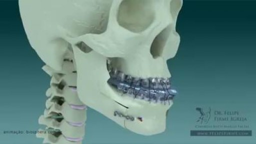
Understanding the process of getting braces

Watch that video of a Snake bite causes girl’s leg to rot away

Orthopedic spine surgeons and vascular surgeons at UW Health in Madison, WI work together to perform minimally invasive anterior lumbar interbody fusion (Mini-ALIF). With this type of spinal fusion surgery, patients have smaller incisions, usually spend less time in the hospital and typically return to daily activities more quickly. Learn more https://www.uwhealth.org/ALIF
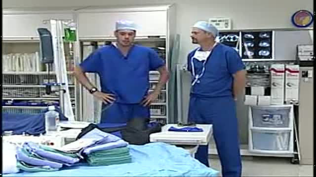
A video showing the accurate steps of Gloving, Gowning and Surgical Scrub
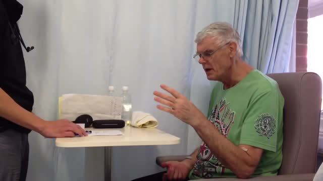
Testing for the four features of Gerstmann Syndrome in this patient with two separate left sided strokes (left frontoparietal ischaemic stroke followed by left posterior parietal haemorrhagic stroke). He exhibits (i) acalculia, (ii) agraphia, (iii) left-right disorientation, and (iv) finger agnosia. Complicating the issue is his obvious nonfluent aphasia (expressive dysphasia) with paraphasic errors (replacing words with associated words (e.g. says 'fork' instead of 'spoon')) and some comprehension issues.

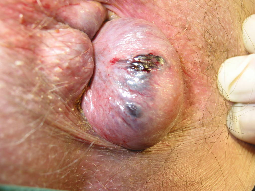
Lichen sclerosus is a skin condition that mainly affects the genital skin (vulva) in women and the penis in men. It most commonly occurs in middle-aged women. Symptoms may include itch, soreness, and changes in the appearance of affected skin.
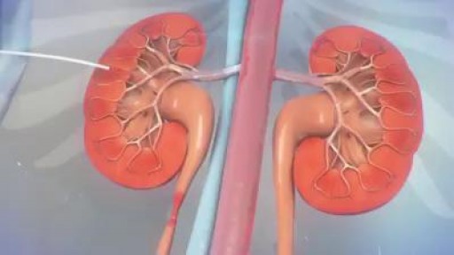
The ureter can become obstructed due to conditions such as kidney stones, tumors, infection, or blood clots. When this happens, physicians can use image guidance to place stents or tubes in the ureter to restore the flow of urine to the bladder. A ureteral stent is a thin, flexible tube threaded into the ureter.

At Nationwide Children’s, our Department of General Pediatric Surgery provides comprehensive surgical care for infants, children and adolescents with congenital and acquired conditions, including major congenital anomalies, traumatic and thermal injuries, and tumors. As the second largest pediatric treatment center in the United States our surgeons perform more than 4,000 operative procedures every year. We are dedicated to clinical excellence, generation of new knowledge through research and the training of the next generation of leaders in children’s surgery. Under the umbrella of a unified program, 11 surgical departments share a common mission, philosophy and approach to patient care.
Pediatric Surgery Program: https://bit.ly/3t4QZef
Pediatric Surgery Fellowship and Residency: https://bit.ly/3qWAWwd
Meet our Pediatric Surgery Team: https://bit.ly/3n39dJh
Fellowship Programs: https://bit.ly/3EX1JNX
Surgical Services: https://bit.ly/3eYDlB8
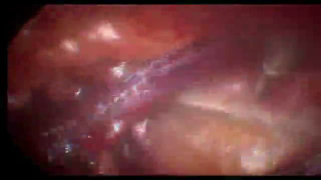
腹腔镜右斜疝修补术+胆囊切除术
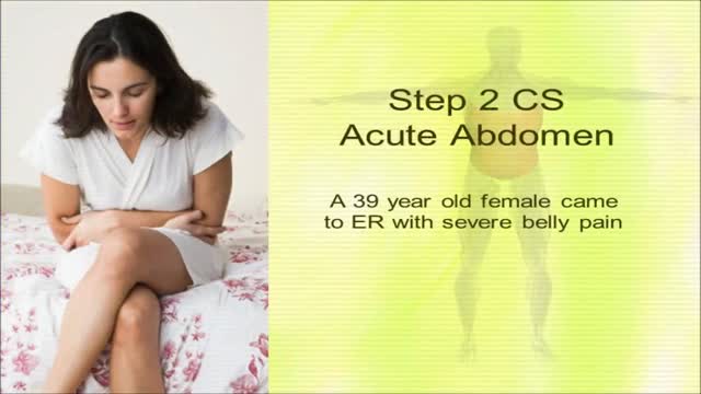
USMLE Step 2 CS - Acute Abdomen- This is just preview video. To get full access please visit our website : www.usmletutoring.com
