- Physical Examination
- Surgical Examination
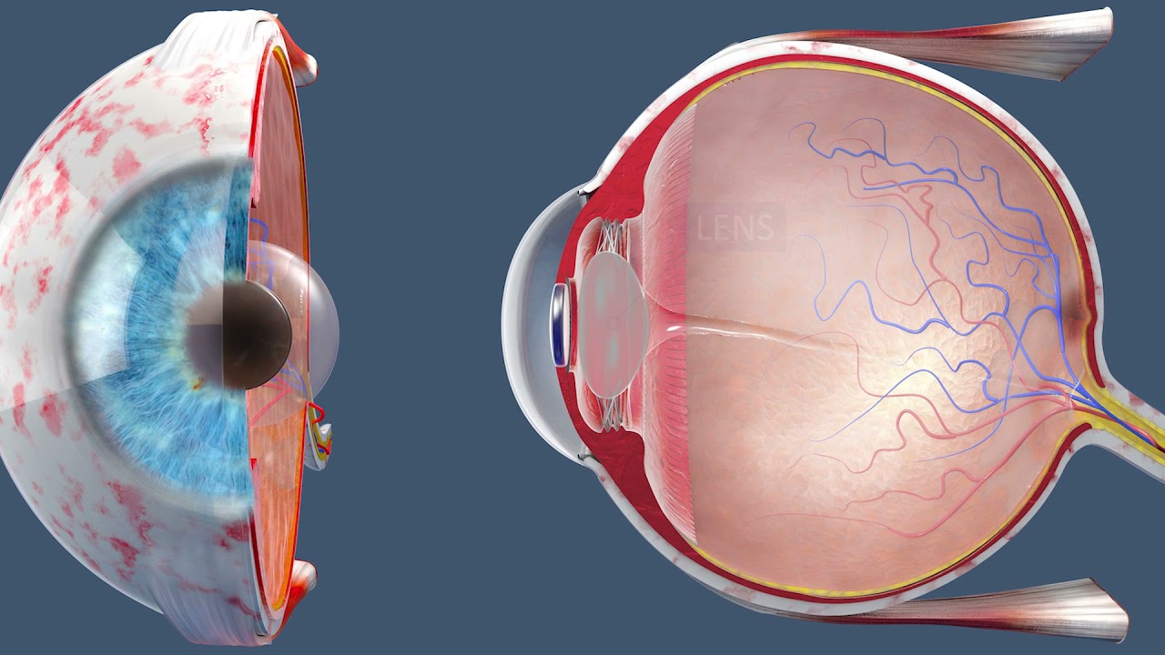
- Ophthalmology
- Clinical Skills
- Orthopedics
- Surgery Videos
- Laparoscopy
- Pediatrics
- Funny Videos
- Cardiothoracic Surgery
- Nursing Videos
- Plastic Surgery
- Otorhinolaryngology
- Histology and Histopathology
- Neurosurgery
- Dermatology
- Pediatric Surgery
- Urology
- Dentistry
- Oncology and Cancers
- Anatomy Videos
- Health and Fitness
- Radiology
- Anaesthesia
- Physical Therapy
- Pharmacology
- Interventional Radiology
- Cardiology
- Endocrinology
- Gynecology
- Emergency Medicine
- Psychiatry and Psychology
- Childbirth Videos
- General Medical Videos
- Nephrology
- Physiology
- Diet and Food Health
- Diabetes Mellitus
- Neurology
- Women Health
- Osteoporosis
- Gastroenterology
- Pulmonology
- Hematology
- Rheumatology
- Toxicology
- Nuclear Medicine
- Infectious Diseases
- Vascular Disease
- Reproductive Health
- Burns and Wound Healing
- Other
Top videos
The patient is awake as a laser cuts her cataract into six pieces. Then, she heads into the operating room. When she wakes up, her cataracts and nearsightedness are gone.
#insidetheor
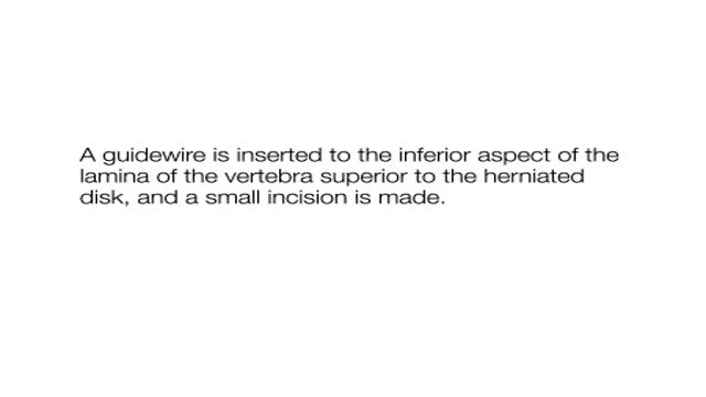
A video showing the procedure of Tubular Diskectomy of a herniated disk. Uploaded on MedicalVideos.us.Discussing the management of Sciatica.
Watch that video of Penile Lengthening and Girth Enhancement Plastic Surgery
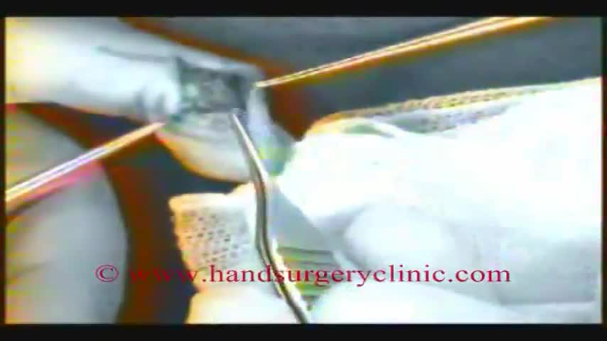
Hernia Repair with Prolene Hernia System

Traumatic penile injury can be due to multiple factors. Penile fracture, penile amputation, penetrating penile injuries, and penile soft tissue injuries are considered urologic emergencies and typically require surgical intervention. The goals of treatment for penile trauma are universal: preservation of penile length, erectile function, and maintenance of the ability to void while standing. Traumatic injury to the penis may concomitantly involve the urethra.[1, 2] Urethral injury and repair is beyond the scope of this article but details can be found in Urethral Trauma. Penile fracture Penile fracture is the traumatic rupture of the corpus cavernosum. Traumatic rupture of the penis is relatively uncommon and is considered a urologic emergency.[3] Sudden blunt trauma or abrupt lateral bending of the penis in an erect state can break the markedly thinned and stiff tunica albuginea, resulting in a fractured penis. One or both corpora may be involved, and concomitant injury to the penile urethra may occur. Urethral trauma is more common when both corpora cavernosa are injured.[4] Penile rupture can usually be diagnosed based solely on history and physical examination findings; however, in equivocal cases, diagnostic cavernosography or MRI should be performed. Concomitant urethral injury must be considered; therefore, preoperative retrograde urethrographic studies should generally be performed. See the images below.
Glomus tumors are rare soft tissue neoplasms that typically present in adults (ages 20-40 years) as small, blue-red papules or nodules of the distal extremities, with most cases involving subungual sites. These tumors are typically painful, often causing paroxysmal pain in response to temperature changes or pressure. Glomus tumors are thought to arise from the glomus body, a thermoregulatory shunt concentrated in the fingers and toes. Most lesions are solitary and localized to cutaneous sites; however, generalized glomuvenous malformations, or multiple glomangiomas, have also been described, and may have extracutaneous involvement.
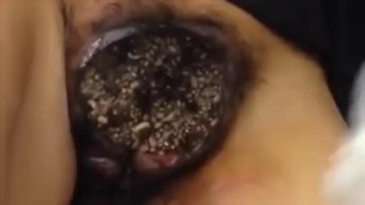
Watch that video of Nasty Female Genital Infection
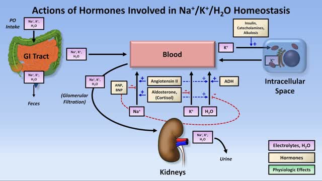
Low potassium (hypokalemia) refers to a lower than normal potassium level in your bloodstream. Potassium is a chemical (electrolyte) that is critical to the proper functioning of nerve and muscles cells, particularly heart muscle cells. Normally, your blood potassium level is 3.6 to 5.2 millimoles per liter (mmol/L). A very low potassium level (less than 2.5 mmol/L) can be life-threatening and requires urgent medical attention.
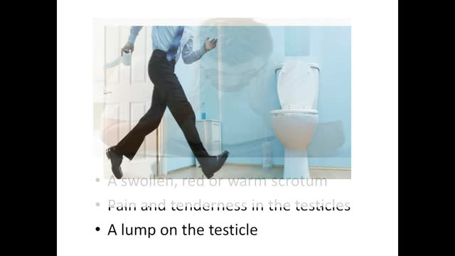
Epididymitis is infection or less frequently, inflammation of the epididymis (the coiled tube on the back of the testicle). The majority of men that develop epididymitis develop it because of a bacterial infection. Although males of any age can develop epididymitis, it occurs most frequently between ages of 20 to 39.
A hernia occurs when an organ or fatty tissue squeezes through a weak spot in a surrounding muscle or connective tissue called fascia. The most common types of hernia are inguinal (inner groin), incisional (resulting from an incision), femoral (outer groin), umbilical (belly button), and hiatal (upper stomach).
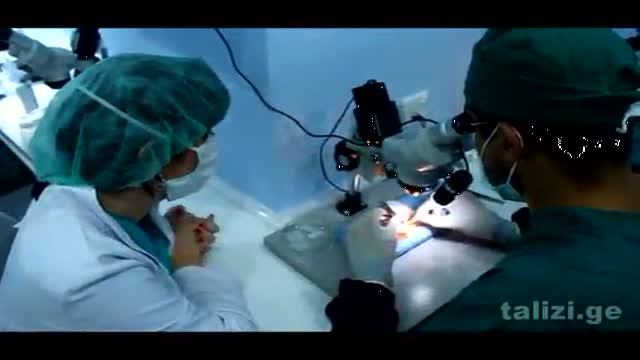
The Talizi Hair Transplantation Clinic offers hair restoration through a painless hair transplantation procedure and guarantees a natural result for an affordable price. 6000 grafts at one session. Hair transplantation surgery combining seamless Follicular Unit Extraction FUE method and Strip Version.

Your egg usually lives for just 12 to 24 hours, but sperm will live inside you for anything from a few hours to seven days, with one to three days the optimum time. ... But because a small number of sperm are long-living, having sex up to six days before ovulation can also result in pregnancy.
Comprehensive physical examination
Marfan syndrome is a disorder of connective tissue, the tissue that strengthens the body's structures. Disorders of connective tissue affect the skeletal system, cardiovascular system, eyes, and skin.
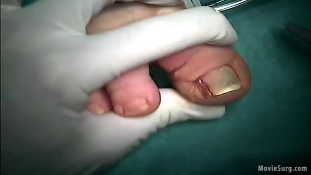
irregular, curved toenails. footwear that places a lot of pressure on the big toes, such as socks and stockings that are too tight or shoes that are too tight, narrow, or flat for your feet. toenail injury, including stubbing your toe, dropping something heavy on your foot, or kicking a ball repeatedly. poor posture.

Bipolar disorder, also known as manic-depressive illness, is a brain disorder that causes unusual shifts in mood, energy, activity levels, and the ability to carry out day-to-day tasks. There are four basic types of bipolar disorder; all of them involve clear changes in mood, energy, and activity levels.

Pigmentflecken, Vitiligo Komplett Geheilt, Pigmentflecken Oberlippe, Vitiligo Heilung, Vitiligo--- http://vitiligo-heilung.info-pro.co/ --- Was ist Vitiligo? Vitiligo ist ein medizinischer Zustand, der die Haut befällt. Die Haut entwickelt dabei an unterschiedlichen Körperstellen nach und nach weiße Flecken, weshalb die Hautstörung im Deutschen auch als "Weißfleckenkrankheit" bekannt ist. Vitiligo macht keine Unterschiede bei Geschlecht oder Rasse und kann jeden befallen. Es wird geschätzt, dass gegenwärtig mehr als 100 Millionen Menschen weltweit in mehr oder minderem starken Maße an Vitiligo leiden. In den Vereinigten Staaten ist die Prävalenzrate etwa 1 % der Bevölkerung; Europa weist ähnliche Raten auf. Was verursacht den Zustand? Vitiligo tritt auf, wenn die Melanozyten in der Haut aus irgendeinem Grund zerstört werden oder ihre Funktion einstellen. Als Melanozyten bezeichnet man jene Hautzellen, die für die Hautfarbe verantwortlich sind. Werden sie zerstört oder anderweitig kompromittiert, stellen produzieren sie kein Melanin mehr, das Hautpigment. Das führt dann zu weißen Hautflecken. Es gibt verschiedene Faktoren, welche die Melanozyten zerstören oder beeinträchtigen können. Dennoch bleibt die Ursache bei den meisten Fällen von Vitiligo ungeklärt. Es wird außerdem angenommen, dass es sich bei Vitiligo um eine Autoimmunerkrankung handelt, welche das Immunsystem die Melanozyten angreifen lässt. Vitiligo kann zudem das Resultat einer Melanozytenstörung sein, welche diese Zellen praktisch veranlasst, "Sebstmord" zu begehen. Laut mancher Forscher kann Vitiligo aber auch durch chronischen Stress und Sonnenbrand hervorgerufen werden. Was sind die Symptome? Das offensichtlichste Symptom von Vitiligo sind die weißen Hautflecken, die sich im Laufe der Zeit vergrößern und auch weiter ausbreiten können. Die Rate, mit der die Hautstörung voranschreitet, unterscheidet sich dabei von Patient zu Patient. "Gratis-Präsentation enthüllt einen ziemlich ungewöhnlichen Tipp zur Beseitigung von Vitiligo für alle Zeiten und in nur 45-60 Tagen - Garantiert!" http://vitiligo-heilung.info-pro.co

Mastitis is inflammation of tissue in one or both mammary glands inside the breast. Mastitis usually affects lactating women - women who are breastfeeding, producing milk. Hence, it is often referred to as lactation mastitis. The patient feels a hard, sore spot inside the breast.
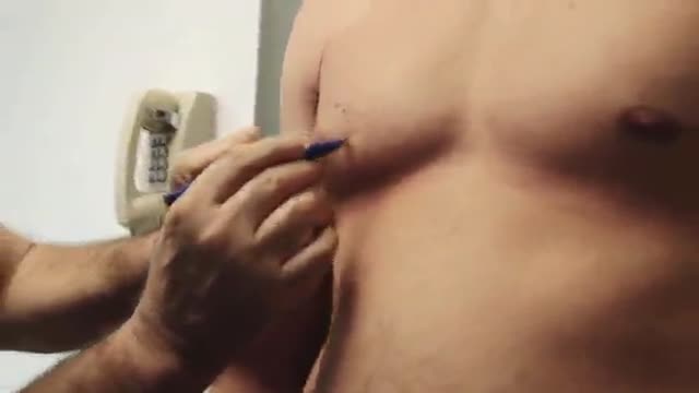
HD Gynecomastia Surgery
How do you know if you have pneumonia? They may include: Cough. You will likely cough up mucus (sputum) from your lungs. ... Fever. Fast breathing and feeling short of breath. Shaking and "teeth-chattering" chills. Chest pain that often feels worse when you cough or breathe in. Fast heartbeat. Feeling very tired or very weak. Nausea and vomiting.
