- Physical Examination
- Surgical Examination
- Ophthalmology
- Clinical Skills
- Orthopedics
- Surgery Videos
- Laparoscopy
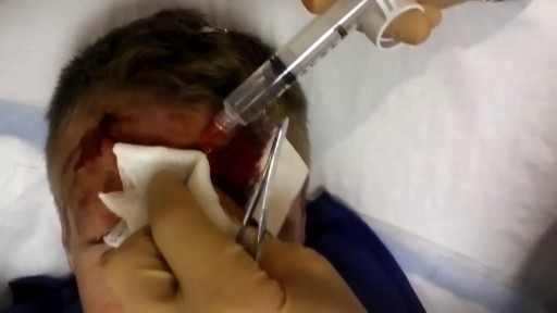
- Pediatrics
- Funny Videos
- Cardiothoracic Surgery
- Nursing Videos
- Plastic Surgery
- Otorhinolaryngology
- Histology and Histopathology
- Neurosurgery
- Dermatology
- Pediatric Surgery
- Urology
- Dentistry
- Oncology and Cancers
- Anatomy Videos
- Health and Fitness
- Radiology
- Anaesthesia
- Physical Therapy
- Pharmacology
- Interventional Radiology
- Cardiology
- Endocrinology
- Gynecology
- Emergency Medicine
- Psychiatry and Psychology
- Childbirth Videos
- General Medical Videos
- Nephrology
- Physiology
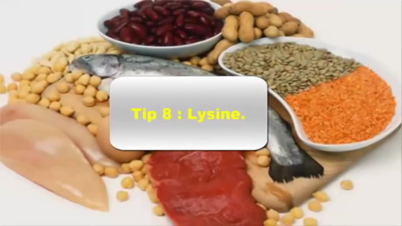
- Diet and Food Health
- Diabetes Mellitus
- Neurology
- Women Health
- Osteoporosis
- Gastroenterology
- Pulmonology
- Hematology
- Rheumatology
- Toxicology
- Nuclear Medicine
- Infectious Diseases
- Vascular Disease
- Reproductive Health
- Burns and Wound Healing
- Other
Top videos
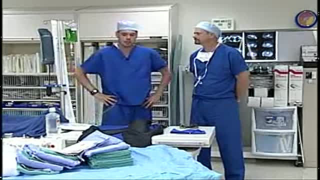
A video showing the accurate steps of Gloving, Gowning and Surgical Scrub
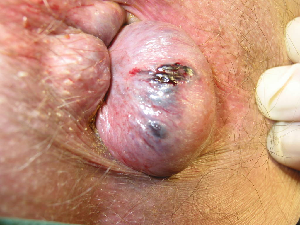
Watch that video to learn everything about Hemorrhoids Repairing Surgery
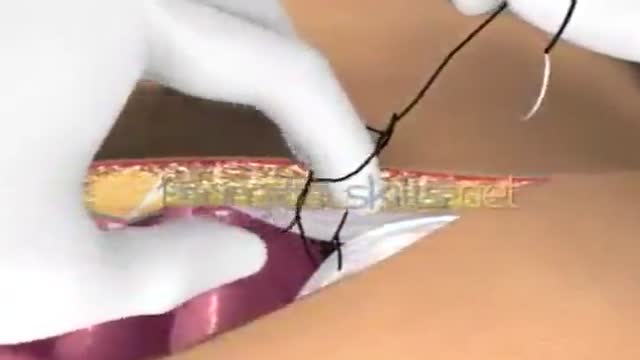
Laparotomy Closure Abdomen Animation
Emergency contraception is a method of birth control you can use if you had sex without using birth control or if your birth control method did not work correctly. You must use emergency contraception as soon as possible after unprotected sex. Emergency contraception pills are different from the abortion pill. If you are already pregnant, emergency contraception pills do not stop or harm your pregnancy. Emergency contraception has also been called the "morning-after pill," but you do not need to wait until the morning after unprotected sex to take it. Emergency contraception is not meant to be used for regular birth control. Talk to your doctor or nurse about regular birth control to help prevent pregnancy. Nearly half of all pregnancies in the United States are unplanned.1
HSV-1 causes "cold sores" on the mouth, and up to 80% of the population has this virus. However, HSV-1 may also be transmitted to the genitals through oral/genital sex and about 40% of genital herpes is caused by HSV-1. Up to 22% of sexually active adults have genital herpes caused by HSV-2.

Watch that video of a Snake bite causes girl’s leg to rot away
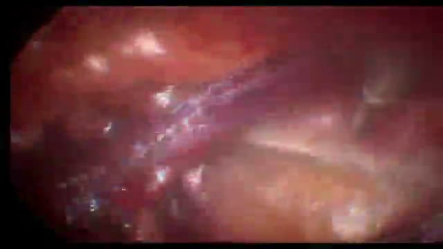
腹腔镜右斜疝修补术+胆囊切除术

Ever heard medical terms like MRI or EKG? Funny speaker for nurses and doctors and all-around healthcare speaker Dr. Brad Nieder discusses the funny medical jargon he's encountered during his medical career.
He jokes about medical acronyms and big healthcare terms. His funny medical humor makes the conference attendees burst with laughter and he reads the medical definition for "laugh."
As an experienced physician and keynote speaker, he's perfect for any in-person or virtual conference or event. He's also a great healthcare speaker to bring in for continuing medical education (cme) units!
Learn more about Brad's keynote and virtual speaking, and book him for your next conference or virtual event: https://www.HealthyHumorist.com
Find Dr. Brad on social media:
https://www.facebook.com/HealthyHumor...
https://www.linkedin.com/in/BradNieder
https://twitter.com/HealthyHumorist
https://www.youtube.com/c/BradNiederMD
https://vimeo.com/BradNieder
Brad Nieder, MD, CSP*
The Healthy Humorist
Doctor, Keynote Speaker, Clean Comedian
*CSP=Certified Speaking Professional
"Medical Lingo"
From the DVD "The Healthy Humorist in Orlando: Laughter is the Best Medicine"
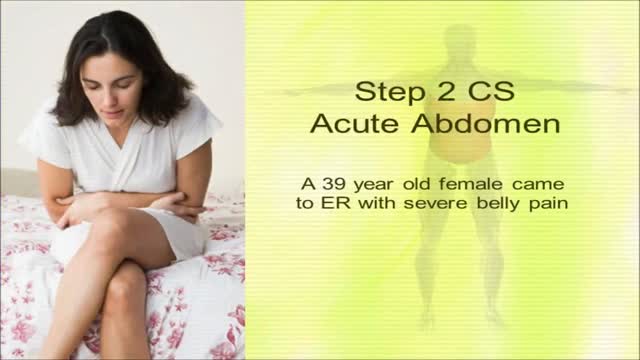
USMLE Step 2 CS - Acute Abdomen- This is just preview video. To get full access please visit our website : www.usmletutoring.com
Possible causes include a combination of biological, psychological, and social sources of distress. Increasingly, research suggests these factors may cause changes in brain function, including altered activity of certain neural circuits in the brain. The persistent feeling of sadness or loss of interest that characterizes major depression can lead to a range of behavioral and physical symptoms. These may include changes in sleep, appetite, energy level, concentration, daily behavior, or self-esteem. Depression can also be associated with thoughts of suicide. The mainstay of treatment is usually medication, talk therapy, or a combination of the two. Increasingly, research suggests these treatments may normalize brain changes associated with depression.
Purchase a license to download a non-watermarked copy of this video here: https://www.alilamedicalmedia.....com/-/galleries/all-
Voice by: Sue Stern.
©Alila Medical Media. All rights reserved.
Support us on Patreon and get FREE downloads and other great rewards: patreon.com/AlilaMedicalMedia
Perfect for patient education purposes.
All images/videos by Alila Medical Media are for information purposes ONLY and are NOT intended to replace professional medical advice, diagnosis or treatment. Always seek the advice of a qualified healthcare provider with any questions you may have regarding a medical condition.
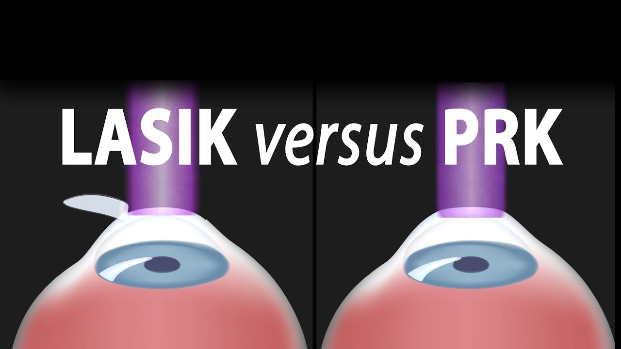
LASIK, or "laser-assisted in situ keratomileusis," is the most commonly performed laser eye surgery to treat myopia, hyperopia and astigmatism. The goal of the treatment is to reshape the cornea to correct the refractive error of the eye.
The cornea is the transparent dome-shaped structure in front of the eye. The cornea refracts light and accounts for about two-thirds of the eye's total optical power. Altering the curvature of the cornea changes the way light rays enter the eye. As a result, the light rays can be focused properly onto the retina for clearer vision.
For nearsighted people, the laser is used to flatten the cornea. For farsighted people, the cornea is made steeper. For patients with astigmatism, the laser is used to smooth the irregularly-shaped cornea into a more regular shape.
The outer layer of the cornea - the epithelium – is capable of replacing itself within a few days after being damaged or removed. The deeper layer of the cornea – the stroma, on the contrary, is a permanent corneal tissue with very limited regenerative capacity. The stroma, if reshaped by a laser, will remain that way permanently.
In this procedure, a thin, circular "FLAP" is created in the surface of the cornea to gain access to the permanent corneal tissue. This can be done with a mechanical cutting tool called a microkeratome, OR, for a blade-free experience, by a femtosecond laser. An excimer laser is then used to remove some corneal tissue to reshape the cornea. Excimer laser uses cool ultraviolet light beams to vaporize microscopic amounts of tissue in a precise manner to accurately reshape the cornea. The excimer laser is computer-controlled and is programmed based on the patient’s refractive error. The flap is then laid back in place and is allowed to heal.
LASIK eye surgery is mostly painless and can be completed within minutes. Improved vision can usually be seen overnight.
PRK, or photorefractive keratectomy, was the first type of laser eye surgery for vision correction and is the predecessor to the popular LASIK procedure. In PRK, NO flap is created. Rather, the epithelial cells on the eye surface are simply removed. An excimer laser is then used to reshape the cornea just like it does in LASIK.
The vision correction outcomes of PRK surgery are comparable to those of LASIK, but the recovery period is longer. This is because the epithelium is completely removed in PRK and it takes a few days to regenerate. PRK patients also have more discomfort and haziness of vision in the first few days after the surgery. Improved vision also takes longer to achieve.
PRK does, however, offer certain advantages. Because PRK does not involve creation of a flap, which contains both epithelial and deeper stromal tissue, the entire thickness of the stroma is available for treatment. The treatment range is therefore higher. This is particularly useful for patients with high levels of myopia or for those whose cornea is too thin for LASIK. PRK is also free of flap-related complication risks.

Endoscopy of Mammary Ducts with Micro-Endoscope called Mammary Ductoscopy. Indication:- Nipple Discharge. In this case Papilloma seen quite clearly. Biopsy can also be possible with Ductoscopy. Mammary Ductoscopy is very useful for diagnosis of Breast Cancer in early stage.
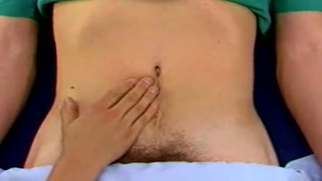
Palpation for Abdominal Masses

Mastitis is inflammation of tissue in one or both mammary glands inside the breast. Mastitis usually affects lactating women - women who are breastfeeding, producing milk. Hence, it is often referred to as lactation mastitis. The patient feels a hard, sore spot inside the breast.

Menopause is the end of menstruation. In clinical terms, you reach menopause when you haven't had a period for 12 months. Vaginal bleeding after menopause isn't normal and should be evaluated by your doctor. For instance, postmenopausal vaginal bleeding can be caused by: Cancer of the uterus, including endometrial cancer and uterine sarcoma Cancer of the cervix or vagina Thinning of the tissues lining the uterus (endometrial atrophy) or vagina (vaginal atrophy) Uterine fibroids Uterine polyps Infection of the uterine lining (endometritis) Medications such as hormone therapy and tamoxifen Pelvic trauma Bleeding from the urinary tract or rectum Excessive overgrowth of the cells that make up the lining of the uterus (endometrial hyperplasia) The cause of your bleeding may be entirely harmless. However, postmenopausal bleeding could result from something serious, so it's important to see your doctor promptly.
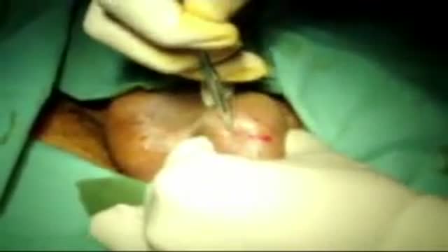
open multi puncture testicular biopsy to retrieve sperm for ICSI (IntaCytoplasmic Sperm Injection) Procedure video
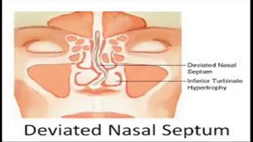
When a deviated septum is severe, it can block one side of your nose and reduce airflow, causing difficulty breathing. The additional exposure of a deviated septum to the drying effect of airflow through the nose may sometimes contribute to crusting or bleeding in certain individuals. Nasal obstruction can occur from a deviated nasal septum, from swelling of the tissues lining the nose, or from both. Treatment of nasal obstruction may include medications to reduce the swelling or nasal dilators that help open the nasal passages. To correct a deviated septum, surgery is necessar
Warning! Do not watch if you are squeamish! SHOW MORE
Masturbating is totally healthy, and totally normal. There are tons of myths out there meant to scare you into thinking masturbation is wrong or bad. But the truth is masturbation is perfectly safe. Masturbating won't make you blind, crazy, or stupid. It won’t damage your genitals, cause pimples, or stunt your growth. It doesn’t use up all your orgasms or ruin other kinds of sex. In fact, masturbation can actually be good for you. Here are some benefits of masturbation: Masturbation is safer than any other type of sex. You can’t get pregnant or get any sexually transmitted infections from masturbating. Masturbation can help you learn what you like and don’t like sexually. And if you decide to have sex with someone, you can know what you do/don’t want to do. BONUS: getting comfortable talking about sex and your body with your partner makes it easier to talk about protecting yourself against STDs and pregnancy, too. Exploring your body and learning how to give yourself sexual pleasure can be empowering and help improve your body image. Masturbation can lower stress and help you relax. It even helps some people fall asleep. Having an orgasm releases endorphins — feel good chemicals in your brain. Orgasms can be a natural painkiller and can even help with period cramps. Mutual masturbation (masturbating with a partner) is a really safe way to have sex and let the other person know what feels good to you. If you share a sex toy, use condoms on the toy and clean it before swapping. And if you touch each other’s genitals, wash your hands before touching your own. Can I get an STD from masturbating? Nope. Masturbating is the safest sexual activity out there. There is virtually NO chance of getting an STD or any other infection from touching your own genitals (and there’s also no chance of pregnancy). STDs have to be passed from one person to another, so you can’t give yourself an STD. The one exception to this is herpes - so if you have any cold sores on your mouth and touch them, make sure to wash your hands before masturbating. But it IS possible to get an STD if you’re masturbating with another person and touching each other’s genitals. Anytime semen (cum) or vaginal fluids are spread to someone else’s body, or your genitals rub against each other, there’s a risk of STDs. So if you touch each other’s genitals, wash your hands before touching your own. STDs can also be spread by sharing sex toys with another person. You can help protect yourself by using condoms on any toys that you share (even if they’re not shaped like a penis). Put a new condom on anytime a different person uses it. If you’re the only one using your sex toys, you don’t have to worry about STDs. But if you use them with other people, protect those sex toys just like you’d protect your own genitals — put a condom on ‘em! It’s possible for masturbation to cause irritation or infections if your body is sensitive to the way you masturbate or the things you masturbate with — but this isn’t the same thing as an STD. Lotions, Vaseline, oils, and scented or flavored stuff may irritate your vulva and vagina. Masturbating roughly and not using lubrication can also lead to irritation because of friction. And germs from the anus can cause vaginal infections — so never put something in your vagina that’s been in your butt without washing it or covering it with a condom. If you’re worried that you have an STD because of pain, itching, or discomfort in your genitals, go to your doctor or your local Planned Parenthood health center.

http://without-glasses.good-info.co How To Improve Eyesight Naturally With Food , How To Improve Eyesight Naturally With Exercises Food. Naturally PERFECT your Vision to 20/20 If you are one of the millions of Americans who suffer from visual problems such as Myopia and Hyperopia then this video will SHOCK you! In the following free video you'll discover: How you can 100% naturally and safely cure almost any visual problem. Why your glasses and contacts are in fact WORSENING your eye condition. The real TRUTH about the Eyecare industry This revolutionary program that you'll soon discover has dared to challenge the billion-dollar Eyecare industry. It reveals this amazing secret to getting 20/20 vision and it doesn't matter what your eye problems are, whether short or long sightedness, Presbyopia, Glaucoma.. whatever! It should help with everything. more information in. http://without-glasses.good-info.co
