- Physical Examination
- Surgical Examination
- Ophthalmology
- Clinical Skills
- Orthopedics
- Surgery Videos
- Laparoscopy
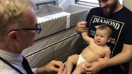
- Pediatrics
- Funny Videos
- Cardiothoracic Surgery
- Nursing Videos
- Plastic Surgery
- Otorhinolaryngology
- Histology and Histopathology
- Neurosurgery
- Dermatology
- Pediatric Surgery
- Urology
- Dentistry
- Oncology and Cancers
- Anatomy Videos
- Health and Fitness
- Radiology
- Anaesthesia
- Physical Therapy
- Pharmacology
- Interventional Radiology
- Cardiology
- Endocrinology
- Gynecology
- Emergency Medicine
- Psychiatry and Psychology
- Childbirth Videos
- General Medical Videos
- Nephrology
- Physiology
- Diet and Food Health
- Diabetes Mellitus
- Neurology
- Women Health
- Osteoporosis
- Gastroenterology
- Pulmonology
- Hematology
- Rheumatology
- Toxicology
- Nuclear Medicine
- Infectious Diseases
- Vascular Disease
- Reproductive Health
- Burns and Wound Healing
- Other
Top videos

Rheum is made up of mucus, skin cells, oils and dust. The rheum that comes from the eyes and forms eye boogers is called gound, which you may know as eye sand, eye gunk, sleep dust, sleep sand, sleep in your eyes, or eye shnooters. When you're awake, gound doesn't cause any problems.
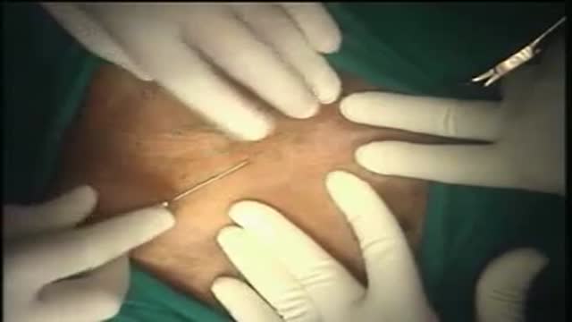
Hernia Repair with Prolene Hernia System
The cornea occupies the front center part of the outer wall of the eye. It is made of collagen fibers in a very special arrangement so that the cornea is clear. One looks through the cornea to see the iris and pupil. The cornea bends light coming into the eye so that it is focused on the retina.
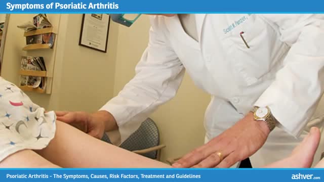
Psoriatic arthritis is a chronic arthritis. In some people, it is mild, with just occasional flare ups. In other people, it is continuous and can cause joint damage if it is not treated. Early diagnosis is important to avoid damage to joints. Psoriatic arthritis typically occurs in people with skin psoriasis, but it can occur in people without skin psoriasis, particularly in those who have relatives with psoriasis. Psoriatic arthritis typically affects the large joints, especially those of the lower extremities, distal joints of the fingers and toes, and also can affect the back and sacroiliac joints of the pelvis. For most people, appropriate treatments will relieve pain, protect the joints, and maintain mobility. Physical activity helps maintain joint movement. Psoriatic arthritis is sometimes misdiagnosed as gout, rheumatoid arthritis or osteoarthritis. - See more at: http://www.rheumatology.org/I-Am-A/Patient-Caregiver/Diseases-Conditions/Psoriatic-Arthritis#sthash.VsBTUw76.dpuf
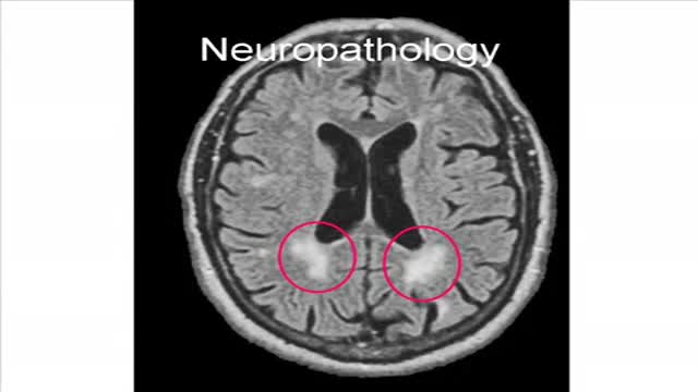
Binswanger's disease is a type of vascular dementia that involves white matter infarcts. Patients with this disease usually present with apathy, agitation, and bilateral corticospinal or bulbar signs
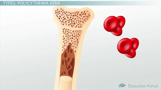
Polycythemia vera (pol-e-sigh-THEE-me-uh VEER-uh) is a slow-growing type of blood cancer in which your bone marrow makes too many red blood cells. Polycythemia vera may also result in production of too many of the other types of blood cells — white blood cells and platelets. These excess cells thicken your blood and cause complications, such as such as a risk of blood clots or bleeding. Polycythemia vera isn't common. It usually develops slowly, and you may have it for years without noticing signs or symptoms. Often, polycythemia vera is found during a blood test done for some other reason. Without treatment, polycythemia vera can be life-threatening. However, with proper medical care, many people experience few problems related to this disease. Over time, there's a risk of progressing to more-serious blood cancers, such as myelofibrosis or acute leukemia.
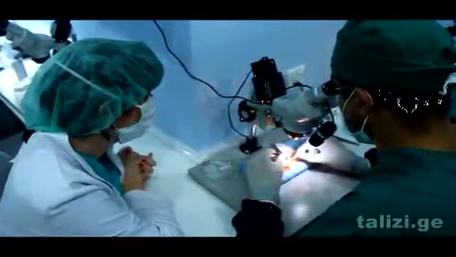
The Talizi Hair Transplantation Clinic offers hair restoration through a painless hair transplantation procedure and guarantees a natural result for an affordable price. 6000 grafts at one session. Hair transplantation surgery combining seamless Follicular Unit Extraction FUE method and Strip Version.
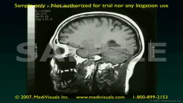
Traumatic brain injury (TBI) is a nondegenerative, noncongenital insult to the brain from an external mechanical force, possibly leading to permanent or temporary impairment of cognitive, physical, and psychosocial functions, with an associated diminished or altered state of consciousness.
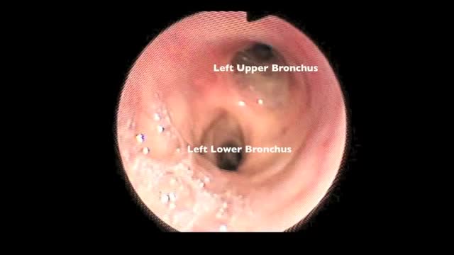
Flexible bronchoscopy is a procedure that allows a clinician to examine the breathing passages (airways) of the lungs (figure 1). Flexible bronchoscopy can be either a diagnostic procedure (to find out more about a possible problem) or a therapeutic procedure (to try to treat an existing problem or condition).
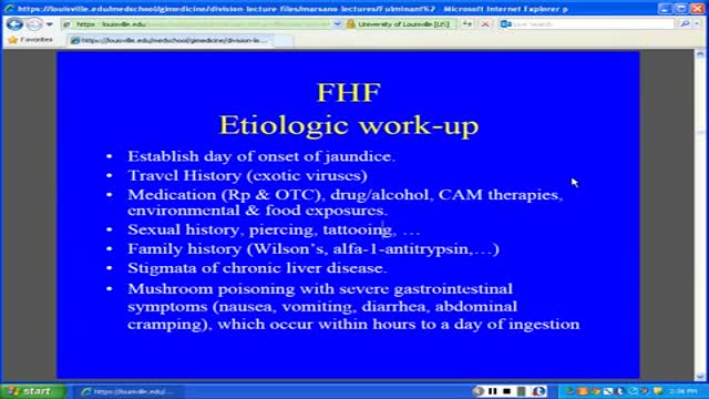
Fulminant hepatic failure (FHF) or acute liver failure (ALF) is defined as the rapid development of acute liver injury with severe impairment of the synthetic function and hepatic encephalopathy in a patient without obvious, previous liver disease.
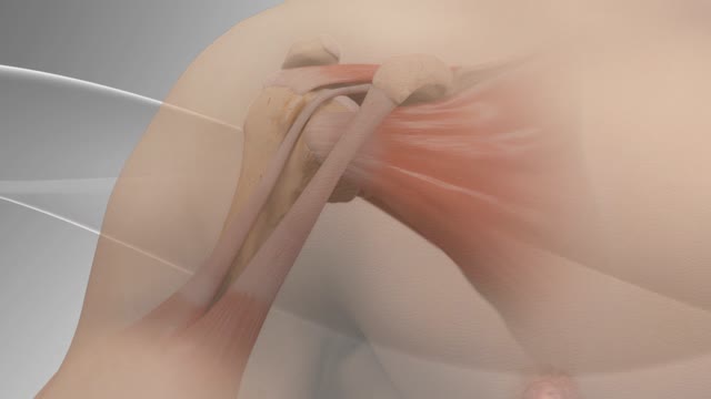
Biceps tenodesis surgery is performed when the biceps tendon is damaged, or the rotator cuff tendon or cartilage ring in the shoulder is torn. The biceps tendon is a strong rope‐like structure connecting the upper end of the biceps muscle to the bones in the shoulder. In biceps tenodesis surgery, the biceps tendon is separated from the shoulder and reattached to the humerus, or the upper arm bone.
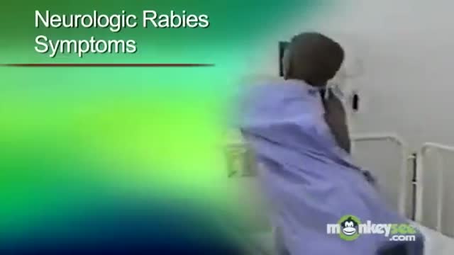
In developing countries, domestic animals (eg, dogs) are common sources of infection. In the United States, bats and wild animals (eg, raccoons) are the most common reservoirs of infection. The acquisition of rabies from bats can occur from an unrecognized bite or a scratch, and possibly by inhalation of aerosolized viral particles. Bats are found in all states except Hawaii, and spelunking (cave exploration) is a risk factor for rabies acquisition from bats.

Each year in the United States, about 400 children and teens younger than age 20 are diagnosed. Osteosarcoma is the third most common cancer in teens, after lymphomas and brain tumors. It is extremely rare in children before age 5.
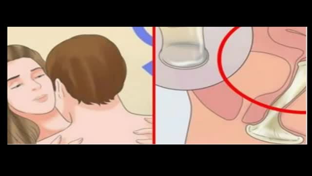
PEOPLE WITH A SEXUALLY ACTIVE LIFE SHOULD BE AWARE OF THIS SILENT KILLER
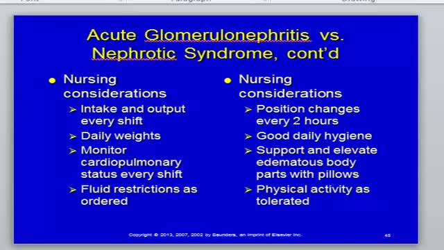
Nephritis and Nephrotic Syndrome
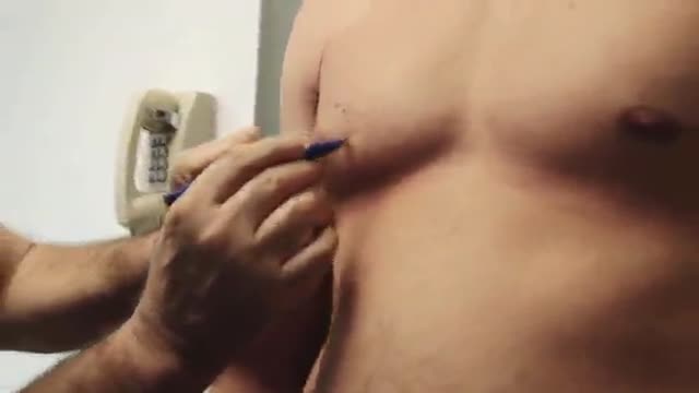
HD Gynecomastia Surgery
Claudication is pain caused by too little blood flow, usually during exercise. Sometimes called intermittent claudication, this condition generally affects the blood vessels in the legs, but claudication can affect the arms, too. At first, you'll probably notice the pain only when you're exercising, but as claudication worsens, the pain may affect you even when you're at rest. Although it's sometimes considered a disease, claudication is technically a symptom of a disease. Most often, claudication is a symptom of peripheral artery disease, a potentially serious but treatable circulation problem in which the vessels that supply blood flow to your legs or arms are narrowed. Fortunately, with treatment, you may be able to maintain an active lifestyle without pain.
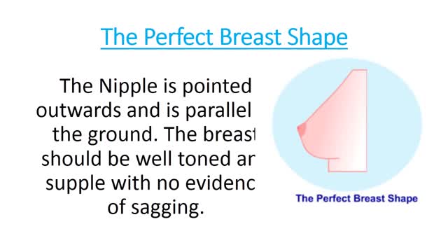
Different Types of Breasts
