- Physical Examination
- Surgical Examination
- Ophthalmology
- Clinical Skills
- Orthopedics
- Surgery Videos
- Laparoscopy
- Pediatrics
- Funny Videos
- Cardiothoracic Surgery
- Nursing Videos
- Plastic Surgery
- Otorhinolaryngology
- Histology and Histopathology
- Neurosurgery
- Dermatology
- Pediatric Surgery
- Urology
- Dentistry
- Oncology and Cancers
- Anatomy Videos
- Health and Fitness
- Radiology
- Anaesthesia
- Physical Therapy
- Pharmacology
- Interventional Radiology
- Cardiology
- Endocrinology
- Gynecology
- Emergency Medicine
- Psychiatry and Psychology
- Childbirth Videos
- General Medical Videos
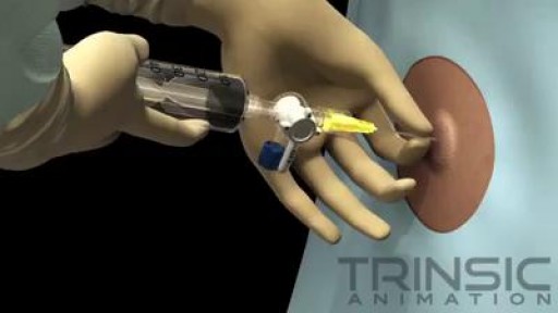
- Nephrology
- Physiology
- Diet and Food Health
- Diabetes Mellitus
- Neurology
- Women Health
- Osteoporosis
- Gastroenterology
- Pulmonology
- Hematology
- Rheumatology
- Toxicology
- Nuclear Medicine
- Infectious Diseases
- Vascular Disease
- Reproductive Health
- Burns and Wound Healing
- Other
Top videos
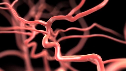
Narrated animation of stroke intervention. Video supplied by Covidien, showing the Solitaire mechanical thrombectomy device, which was the first FDA-approved device for such an indication.
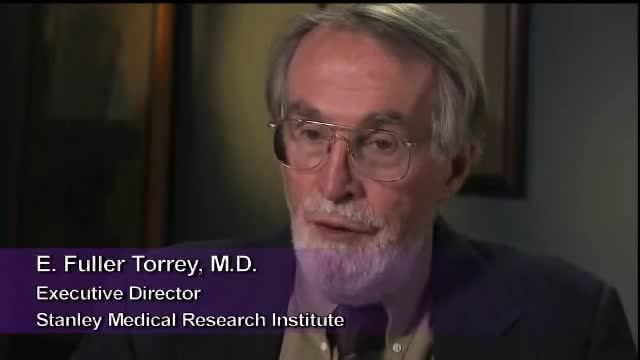
Impaired awareness of illness (anosognosia) is a major problem because it is the single largest reason why individuals with schizophrenia and bipolar disorder do not take their medications. It is caused by damage to specific parts of the brain, especially the right hemisphere.
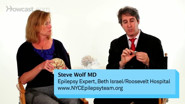
Surgery is an elective procedure done in people who have had extensive testing to decide if they are potential candidates. The following criteria are considered when determining if a person may be a good candidate for surgery. Person has failed adequate trials of two first-line seizure medicines (ones that are commonly effective in controlling the type of seizures the person is experiencing) and one combination of at least two drugs. A trial of a medication is considered adequate when it has been increased gradually to the maximum dosage that does not cause serious side effects. If the person has frequent seizures, any improvement will be obvious after a short time. If the seizures generally occur far apart, however, it may take months to determine whether a medication is helping. At some epilepsy centers, patients are offered additional conventional or experimental medications before surgery is considered. But research suggests that each time a trial of medication fails to control a person's seizures, it becomes less likely that a different medicine or combination will be successful. Since uncontrolled seizures present serious physical risks and social and psychological consequences, the trend these days is to proceed with surgery much sooner than in the past if it seems appropriate for that person.

Having surgery can be frightening for anyone, but it's especially scary for kids who don't always understand what's going on, or what the grown-ups are saying. We're here to help!
Join Avrie, who had surgery at the Sacred Heart Children's Hospital pediatric surgery center in Spokane, WA. Maybe after watching and hearing her story, you and your kiddo will feel better about having surgery in the hospital.
Follow Avrie's trip - from check-in, vital signs and pre-op checks; meeting the doctor who will do his surgery, along with the anesthesiologist, surgery nurse and the Child Life Specialist; the trip to the Operating Room; waking up in the recovery room with his mom by his side; and getting ready to go home.
To learn more about the pediatric surgery center at Sacred Heart Children's Hospital, visit https://washington.providence.....org/locations-direct
Less pain and no incisions are just two benefits of robotically assisted surgery thanks to the da Vinci Surgical System. ~ Detroit Medical Center

Cardiac arrest is the abrupt loss of heart function in a person who may or may not have diagnosed heart disease. The time and mode of death are unexpected. It occurs instantly or shortly after symptoms appear. Each year, more than 350,000 emergency medical services-assessed out-of-hospital cardiac arrests occur in the United States
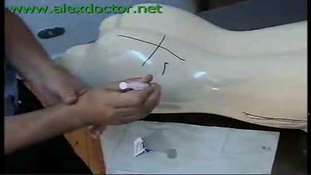
Introducing an IM Injection
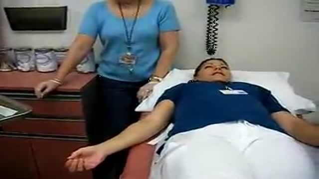
Draw Blood Samples
Thoracentesis is a procedure in which a needle is inserted into the pleural space between the lungs and the chest wall. This procedure is done to remove excess fluid, known as a pleural effusion, from the pleural space to help you breathe easier. It may be done to determine the cause of your pleural effusion. Some conditions such as heart failure, lung infections, and tumors can cause pleural effusions.

The eyes A close up of a young person's eyes. The eyes are responsible for four-fifths of all the information our brain receives. Here you can find out a bit more about how they work, common problems that affect vision and the work Sightsavers does to treat and prevent avoidable blindness. You can also find out more about the people whose lives have been changed thanks to donations from people like you. How do eyes work? (click image to see enlarged version or click here for text alternative) Graphic of an eye with information about its different parts The images we see are made up of light reflected from the objects we look at. This light enters the eye through the cornea. Because this part of the eye is curved, it bends the light, creating an upside down image on the retina (this is eventually put the right way up by the brain). The retina is a complex part of the eye, but only the very back of it is light sensitive. This part of the retina has roughly the area of a 10p coin, and is packed with photosensitive cells called rods and cones. Cones are the cells responsible for daylight vision. There are three kinds – each responding to a different wavelength of light: red, green and blue. The cones allow us to see images in colour and detail. Rods are responsible for night vision. They are sensitive to light but not to colour. In darkness, the cones do not function at all. How do we see an image? The lens focuses the image. It can do this because it is adjustable – using muscles to change shape and help us focus on objects at different distances. The automatic focusing of the lens is a reflex response and is not controlled by the brain. Once the image is clearly focused on the sensitive part of the retina, energy in the light that makes up that image creates an electrical signal. Nerve impulses can then carry information about that image to the brain through the optic nerve.
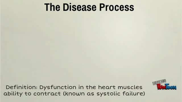
Dilated cardiomyopathy (DCM) is a condition in which the heart's ability to pump blood is decreased because the heart's main pumping chamber, the left ventricle, is enlarged and weakened.

This video documents the experience of one of our Mommy Makeover patients. She is 39 years old, 5’4” tall, and of average weight. Following the birth of her twins, she wanted to improve her abdominal wall contour and correct the lack of shape and firmness in her breasts.

It depends upon which ligament is injured. If it is medial collateral ligament you feel pain when you walk ,sit and stand and you will be liming as well. If it is anterior cruciate ligament you feel pain when you walk on uneven ground.
Phlebotomy Drawing Blood from Veins
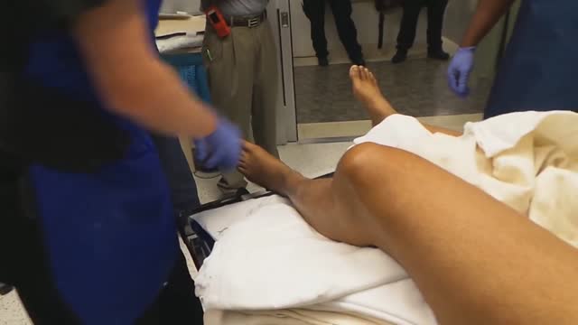
This video has been shortened for quicker review of the procedure. This patient's knee was dislocated during a motor vehicle accident. In this video the reduction of the dislocated knee is demonstrated.

More videos on my youtube channel

Watch that video to know about the Health Benefits from KISSING
The Cardiac Cycle
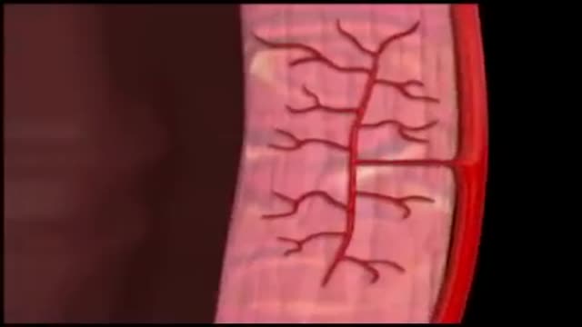
Coronary circulation is the circulation of blood in the blood vessels of the heart muscle (myocardium). The vessels that deliver oxygen-rich blood to the myocardium are known as coronary arteries. The vessels that remove the deoxygenated blood from the heart muscle are known as cardiac veins.
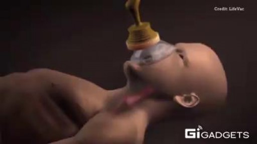
new Anti-Choking Device.
