- Physical Examination
- Surgical Examination
- Ophthalmology
- Clinical Skills
- Orthopedics
- Surgery Videos
- Laparoscopy
- Pediatrics
- Funny Videos
- Cardiothoracic Surgery
- Nursing Videos
- Plastic Surgery
- Otorhinolaryngology
- Histology and Histopathology
- Neurosurgery
- Dermatology
- Pediatric Surgery
- Urology
- Dentistry
- Oncology and Cancers
- Anatomy Videos
- Health and Fitness
- Radiology
- Anaesthesia
- Physical Therapy
- Pharmacology
- Interventional Radiology
- Cardiology
- Endocrinology
- Gynecology
- Emergency Medicine
- Psychiatry and Psychology
- Childbirth Videos
- General Medical Videos
- Nephrology
- Physiology
- Diet and Food Health
- Diabetes Mellitus
- Neurology
- Women Health
- Osteoporosis
- Gastroenterology
- Pulmonology
- Hematology
- Rheumatology
- Toxicology
- Nuclear Medicine
- Infectious Diseases
- Vascular Disease
- Reproductive Health
- Burns and Wound Healing
- Other
Top videos
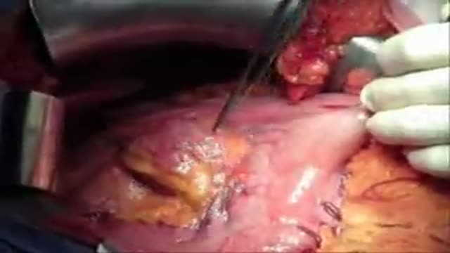
Wedge Resection of a gastric GIST
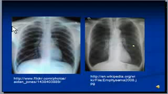
The video will describe radiologic features of Emphysema on a chest x-ray. Please see my website for disclaimer.
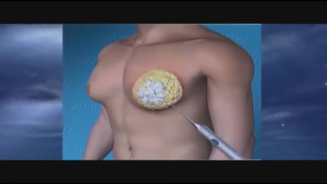
Gynecomastia 3D Animation

#CNA_Practice_Test Welcome to This CNA practice test 15 Basic Nursing Skills Fully Explained Answers. Includes questions from 171 to 180 of These 270 questions that are very similar to the real test #CNA_EXAM.
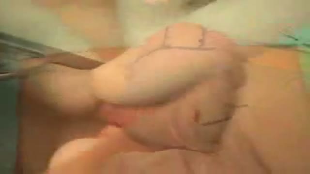
it's the video of the OR during a dynamic reconstruction of the achilles tendon by a composite anterolateral perforator flap
a video showing the technique of Shave and Punch Skin Biopsies nique of
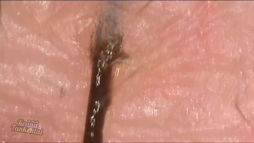
Watch that Ingrown Hair Removal Close up 200X Video

Peptic Ulcer 3D
A video showing squared notch

Function and Anatomy:
Hinge type joint formed by the articulation of the Ulna and Radius (bones of the forearm), and Humerus (upper arm). Full extension is equal to 0 degrees, full flexion to ~ 150 degrees. Maximum supination (turning hand palm up so that it can hold a bowl of "soup") and pronation (palm down) are both 90 degrees

Acetaminophen and ibuprofen are both good medicines, and both provide the same basic relief from fever and pain, even though they have different chemical structures and side effects
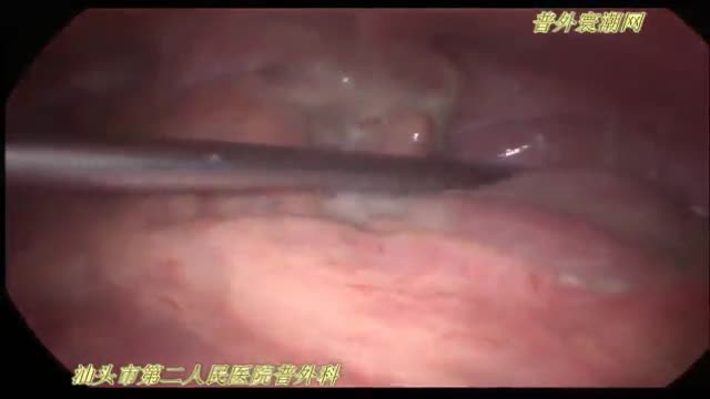
急性坏疽性阑尾炎并穿孔de 腹腔镜手术治疗

Atrial flutter is a type of abnormal heart rate, or arrhythmia. It occurs when the upper chambers of your heart beat too fast. When the chambers in the top of your heart (atria) beat faster than the bottom ones (ventricles), it complicates your heart rhythm

wide resection of giant cell tumor ,then strut grafting using free fibula graft,knowles pinning of the graft.
Distal Urethroplasty with Dorsal Dartos Flap
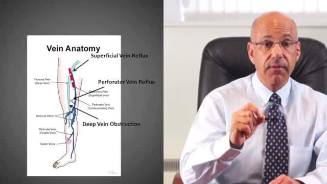
A leg ulcer is simply a break in the skin of the leg, which allows air and bacteria to get into the underlying tissue. This is usually caused by an injury, often a minor one that breaks the skin. In most people such an injury will heal up without difficulty within a week or two. However, when there is an underlying problem the skin does not heal and the area of breakdown can increase in size. This is a chronic leg ulcer.
Multiple sclerosis (MS) involves an immune-mediated process in which an abnormal response of the body’s immune system is directed against the central nervous system (CNS). The CNS is made up of the brain, spinal cord and optic nerves.
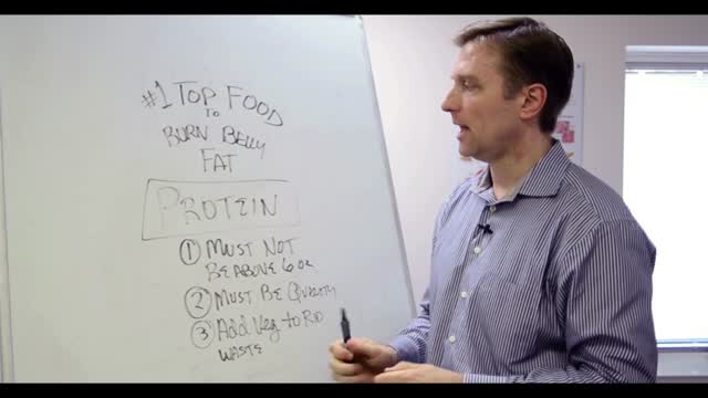
Overweight does not necessarily equal unhealthy. There are actually plenty of overweight people who are in excellent health (1). Conversely, many normal weight people have the metabolic problems associated with obesity (2). That’s because the fat under the skin is actually not that big of a problem (at least not from a health standpoint, it’s more of a cosmetic problem). It’s the fat in the abdominal cavity, the belly fat, that causes the biggest issues (3). If you have a lot of excess fat around your waistline, even if you’re not very heavy, then you should take some steps to get rid of it. Belly fat is usually estimated by measuring the circumference around your waist. This can easily be done at home with a simple tape measure. Anything above 40 inches (102 cm) in men and 35 inches (88 cm) in women, is known as abdominal obesity. There are actually a few proven strategies that have been shown to target the fat in the belly area more than other areas of the body.

Watch that video to know How to Get Pregnant With Twins
We have just enhanced the smile of another wonderful patient! She just received 6 mini dental implants place by DR. Jue www.sugarlanddentalspa.com.
