- Physical Examination
- Surgical Examination
- Ophthalmology
- Clinical Skills
- Orthopedics
- Surgery Videos
- Laparoscopy
- Pediatrics
- Funny Videos
- Cardiothoracic Surgery
- Nursing Videos
- Plastic Surgery
- Otorhinolaryngology
- Histology and Histopathology
- Neurosurgery
- Dermatology
- Pediatric Surgery
- Urology
- Dentistry
- Oncology and Cancers
- Anatomy Videos
- Health and Fitness
- Radiology
- Anaesthesia
- Physical Therapy
- Pharmacology
- Interventional Radiology
- Cardiology
- Endocrinology
- Gynecology
- Emergency Medicine
- Psychiatry and Psychology
- Childbirth Videos
- General Medical Videos
- Nephrology
- Physiology
- Diet and Food Health
- Diabetes Mellitus
- Neurology
- Women Health
- Osteoporosis
- Gastroenterology
- Pulmonology
- Hematology
- Rheumatology
- Toxicology
- Nuclear Medicine
- Infectious Diseases
- Vascular Disease
- Reproductive Health
- Burns and Wound Healing
- Other
Top videos
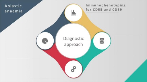
Aplastic anemia is a hematopoietic disorder caused due to T lymphocyte mediated destruction of stem cells resulting in pancytopenia with a cellular bone marrow and normal cell cytogenetics. The causes of aplastic anaemia may be inherited or acquired. The causes and the diagnostic approach, along with spectrum of severity of this disorder is discussed in this presentation. A detailed discussion of the management options, along with pharmacological therapy and supportive therapy in these cases is also discussed. The treatment options include, in addition to a stem cell transplant, anti-thymocyte globulin, cyclosporine, methyprednisolone and eltrombopag (for patients who have failed treatment on combined modality therapy with ATG and cyclosporine)
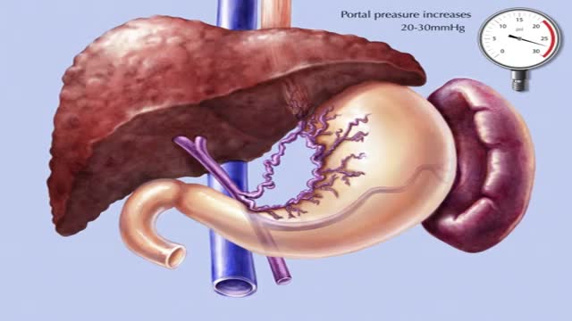
Transjugular intrahepatic portosystemic shunt or transjugular intrahepatic portosystemic stent shunting (commonly abbreviated as TIPS or TIPSS) is an artificial channel within the liver that establishes communication between the inflow portal vein and the outflow hepatic vein.
USMLE Step 2 CS - LGIB This is just preview video. To get full access please visit our website : www.usmletutoring.com
USMLE Step 2 CS - Palpitations This is just preview video. To get full access please visit our website : www.usmletutoring.com

James Slover, MD, and Ivan Madrid, MD, describe the benefits of knee replacement surgery, the differences in partial and total knee replacement, and how the procedures are performed at NYU Langone.
Learn more about Dr. Slover: http://nyulangone.org/doctors/....1851355564/james-d-s
Learn more about Dr. Madrid: http://nyulangone.org/doctors/....1912940107/ivan-madr
To learn more about joint replacement surgery at NYU Langone, visit: http://nyulangone.org/location....s/center-for-musculo
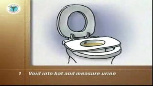
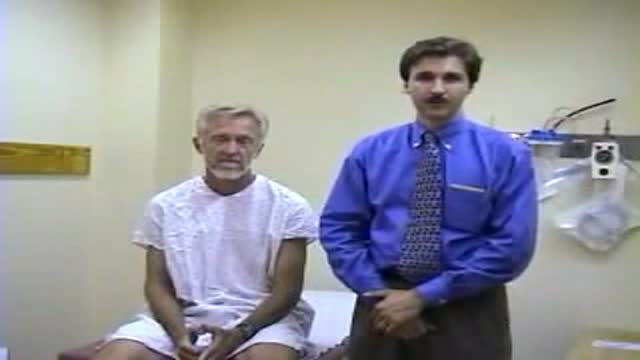
Female Intermittent Self Catheterization
The 30 minute DVD:
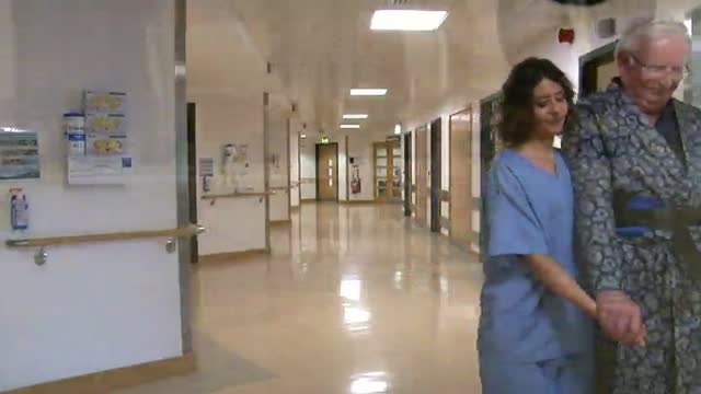
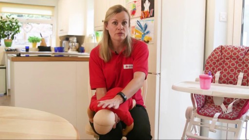
introduces moving and handling of people
describes safer people handling practices
features specialist guidance from a chartered physiotherapist
outlines the process for people handling risk assessments
sets out the principles of safer handling
demonstrates the key safer handling techniques:
rolling a person
inserting and removing sliding sheets
repositioning people using sliding sheets
assisting people to stand and walk with handling belts
the use of roll boards in lateral transfers
using hoists
highlights the important role you play in safer people handling
Myomectomy means the surgical removal of just the fibroid, with reconstruction and repair of the uterus. There are now a number of techniques used to perform myomectomy: through an abdominal incision, vaginal incision, with a laparoscope
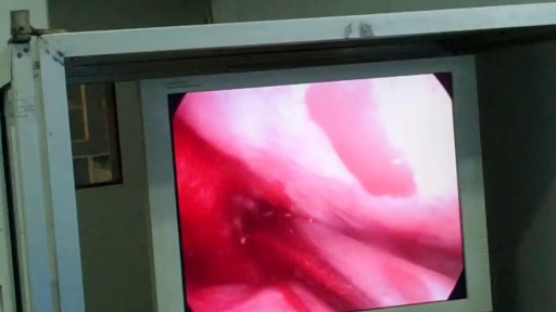
Causes are chronic inflammation due to infection, allergies, drug sensitivity, or immune disorders. Symptoms may include a runny nose, stuffiness, or post-nasal drip. In some cases, there may be no symptoms. The condition can be treated with corticosteroids, other medications, or surgery.
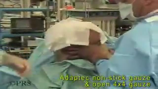
Bandaging a freshly above the knee amputated limb
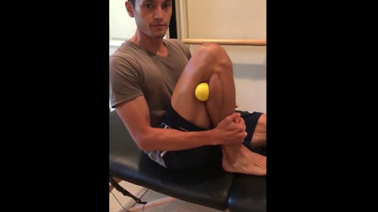
A displaced fibular head can create tightness, pain, and even numbness or tingling along the outside of your knee and down your leg. This most often occurs after a modest hyperextension knee injury, such as landing on one leg after jumping. If you have lingering knee pain and are searching for an answer, try this move
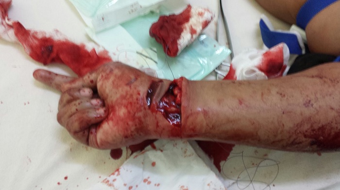
Watch that video to know How to Stop Arterial Bleeding
A new well designed randomized study has suggested that long term baby aspirin usage may aid in fight against cancer. The suggested mechanism is that cancers induce inflammatory responses so the anti-inflammatory mechanism of prostaglandins inhibitors may cease the progress of many cancers. There are some concerns about the study because despite the well-designed randomized study; the study didn't include a satisfying number of female participants. The study was also conducted on esophageal, colorectal and lung cancers.
Vital Signs and Chest Examination

Watch that video to know the Types of Female Genital Infection Yeast or Candidiasis, Trichomoniasis, Bacterial Vaginosis

Best facial cosmetic surgeons Best facial plastic surgeon

Devi Shetty, founder of Narayana Health in India, reflects on the remarkable fact that, after 26 years of operation, the cost of heart surgery at Narayana Health has come down dramatically, and shares some of the strategies used to maintain high quality with low patient cost.
Learn more about the Creating Emerging Markets Project and explore its many compelling interviews: https://www.hbs.edu/creating-e....merging-markets/Page
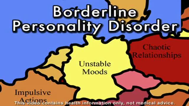
Borderline Personality Disorder Information
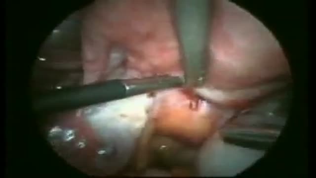
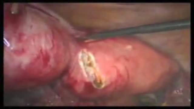
Excision of Rectovaginal Nodule
