- Physical Examination
- Surgical Examination
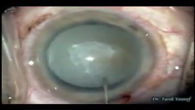
- Ophthalmology
- Clinical Skills
- Orthopedics
- Surgery Videos
- Laparoscopy
- Pediatrics
- Funny Videos
- Cardiothoracic Surgery
- Nursing Videos
- Plastic Surgery
- Otorhinolaryngology
- Histology and Histopathology
- Neurosurgery
- Dermatology
- Pediatric Surgery
- Urology
- Dentistry
- Oncology and Cancers
- Anatomy Videos
- Health and Fitness
- Radiology
- Anaesthesia
- Physical Therapy
- Pharmacology
- Interventional Radiology
- Cardiology
- Endocrinology
- Gynecology
- Emergency Medicine
- Psychiatry and Psychology
- Childbirth Videos
- General Medical Videos
- Nephrology
- Physiology
- Diet and Food Health
- Diabetes Mellitus
- Neurology
- Women Health
- Osteoporosis
- Gastroenterology
- Pulmonology
- Hematology
- Rheumatology
- Toxicology
- Nuclear Medicine
- Infectious Diseases
- Vascular Disease
- Reproductive Health
- Burns and Wound Healing
- Other


Histological staining: hematoxylin & eosin
The most popular and one of the principal stains in histology is hematoxylin and eosin stain. It gives us an overview of the tissue and its structure. Hematoxylin binds with basophilic structures – for example DNA and RNA. So we can observe nuclei stained in blue or purple color. Eosin binds to acidophilic substances such as positively charged amino acid side chains. So as the result cytoplasm is pink or orange. All samples in laboratory are stained with H&E. There are several different types of hematoxylins and eosins used in histology which will give us different results.
In this video you will see, how we stain slides with different types of hematoxylins and eosins. Finally, we will compare the results.
• Subscribe to our channel: https://www.youtube.com/c/BioVitrumEN
• Watch other videos about histological process: https://www.youtube.com/playli....st?list=PLw4LQHit0MU
• Our website: http://en.biovitrum.ru/
