- Physical Examination
- Surgical Examination
- Ophthalmology
- Clinical Skills
- Orthopedics
- Surgery Videos
- Laparoscopy
- Pediatrics
- Funny Videos
- Cardiothoracic Surgery
- Nursing Videos
- Plastic Surgery
- Otorhinolaryngology
- Histology and Histopathology
- Neurosurgery
- Dermatology
- Pediatric Surgery
- Urology
- Dentistry
- Oncology and Cancers
- Anatomy Videos
- Health and Fitness
- Radiology
- Anaesthesia
- Physical Therapy
- Pharmacology
- Interventional Radiology
- Cardiology
- Endocrinology
- Gynecology
- Emergency Medicine
- Psychiatry and Psychology
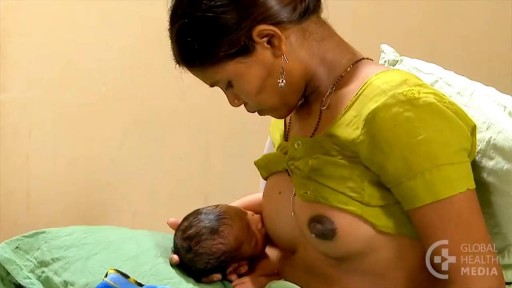
- Childbirth Videos
- General Medical Videos
- Nephrology
- Physiology
- Diet and Food Health
- Diabetes Mellitus
- Neurology
- Women Health
- Osteoporosis
- Gastroenterology
- Pulmonology
- Hematology
- Rheumatology
- Toxicology
- Nuclear Medicine
- Infectious Diseases
- Vascular Disease
- Reproductive Health
- Burns and Wound Healing
- Other


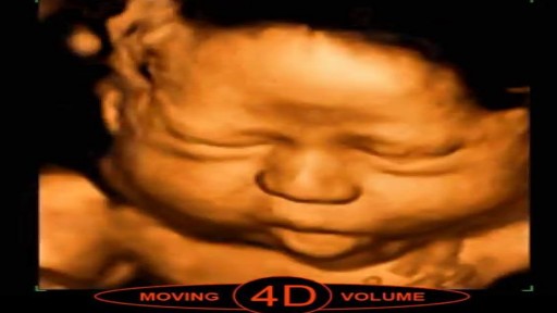


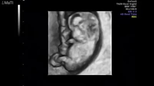
How to use Ultrasound in Pregnancy
A prenatal ultrasound (also called a sonogram) is a noninvasive diagnostic test that uses sound waves to create a visual image of your baby, placenta, and uterus, as well as other pelvic organs. It allows your healthcare practitioner to gather valuable information about the progress of your pregnancy and your baby's health. During the test, an ultrasound technician (sonographer) transmits high-frequency sound waves through your uterus that bounce off your baby. A computer then translates the echoing sounds into video images that reveal your baby's shape, position, and movements. (Ultrasound waves are also used in the handheld instrument called a Doppler that your practitioner uses during your prenatal visits to listen to your baby's heartbeat.) You may have an early ultrasound at your practitioner's office at 6 to 10 weeks to confirm and date the pregnancy. Or you may not have one until the standard midpregnancy ultrasound between 16 and 20 weeks. That's when you may learn your baby's sex, if you like. (The technician will probably present you with a grainy printout of the sonogram as a keepsake.) You may also have a sonogram as part of a genetic test, such as the nuchal translucency test, chorionic villus sampling, or amniocentesis, or at any other time if there are signs of a problem with your baby. You'll have more frequent ultrasounds if you have diabetes, hypertension, or other medical complications.
