- Physical Examination
- Surgical Examination
- Ophthalmology
- Clinical Skills
- Orthopedics
- Surgery Videos
- Laparoscopy
- Pediatrics
- Funny Videos
- Cardiothoracic Surgery
- Nursing Videos
- Plastic Surgery
- Otorhinolaryngology
- Histology and Histopathology
- Neurosurgery
- Dermatology
- Pediatric Surgery
- Urology
- Dentistry
- Oncology and Cancers
- Anatomy Videos
- Health and Fitness
- Radiology
- Anaesthesia
- Physical Therapy
- Pharmacology
- Interventional Radiology
- Cardiology
- Endocrinology
- Gynecology
- Emergency Medicine
- Psychiatry and Psychology
- Childbirth Videos
- General Medical Videos
- Nephrology
- Physiology
- Diet and Food Health
- Diabetes Mellitus
- Neurology
- Women Health
- Osteoporosis
- Gastroenterology
- Pulmonology
- Hematology
- Rheumatology
- Toxicology
- Nuclear Medicine
- Infectious Diseases
- Vascular Disease
- Reproductive Health
- Burns and Wound Healing
- Other


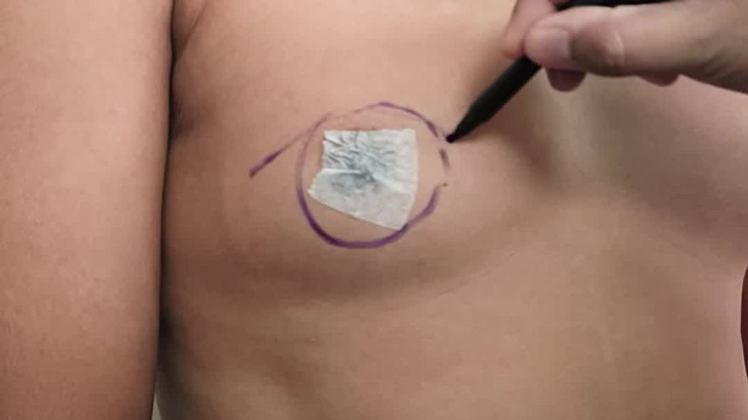


Spina Bifida Surgery Myelomeningocele Repair
What is Myelomeningocele and how does it affect my baby? Myelomeningocele (MMC), one of the most severe forms of spina bifida, is a condition where the fetus’ spinal cord fails to close during development. This happens between 20 and 28 days of gestation, often before a woman knows she is pregnant. Because the spinal cord does not close, many of the nerves are exposed, resulting in damage to the cord as the pregnancy continues. Spina bifida can impact the nervous system, bones and muscles, kidneys and bladder. The location on the spine where the undeveloped area occurs is called the level of the spina bifida. Because nerve damage at this site prevents function below that level, the higher the level, the greater the impact on normal development and function. The opening in the spinal cord also results in loss of the fluid surrounding the nervous system. This causes the brain to be positioned further down into the upper spinal column than normal, which is called an Arnold Chiari II malformation. When this happens, the normal flow of fluid out of the brain is obstructed, causing Hydrocephalus, an excess of cerebrospinal fluid within the brain. After birth, most children with Hydrocephalus need to have the extra fluid shunted out of the brain into the abdomen via a ventriculoperitoneal shunt. MMC affects about 1 in every 1,000 babies, and it ranges in severity. Some children, with mild cases very low on the spinal cord, can function nearly normally. More severe cases can cause leg weakness and paralysis, as well as Hydrocephalus, and the Arnold Chiari malformation. People with MMC often live long lives, especially if the condition is diagnosed and treated early. How is Myelomeningocele diagnosed? At about 15 weeks gestation, a blood test measuring the levels of alpha-fetoprotein can show the physician that there might be a problem. After that, an ultrasound is performed to detect the MMC, but also to detect the conditions that can result from it, such as Hydrocephalus, the Chiari malformation, and any problems with the lower extremities. In all cases, we perform a fetal MRI to gain more detailed information and we perform a fetal echocardiogram (echo) to rule out any problem with the heart. What is the prenatal surgery for Myelomeningocele, and how does it differ from postnatal surgery? Until recently, the only way to treat MMC was surgery after birth. But, now that the nine-year long Management of Myelomeningocele (MOMS) trial has been completed, we know that repairing the MMC before birth, in the womb, can be beneficial to the baby’s outcome. The SSM Health St. Louis Fetal Care Institute has one of the fastest growing fetal MMC repair programs in the country. The results of the trial found that prenatal treatment helps reduce, or even eliminate, the major complications of MMC—the Hydrocephalus, the Chiari malformation, and the lack of movement in the lower extremities. Diagnosis and repair of the MMC before birth can make a big difference in the way the baby develops. The MOMS trial showed that babies treated in the womb need half the VP shunts, often have reversal of the Arnold-Chiari malformation, and are more likely to walk, at least until 30 months. Long-term follow up data of children treated with prenatal surgery is still being collected, so the benefit beyond 30 months is not fully known. The operation for open fetal surgery for MMC repair involves making a small opening in the uterus, then closing the spinal cord opening just like after birth. The womb is repaired and the mother is in the hospital for four to five days. The surgery is performed between 19 and 26 weeks of the pregnancy. Mothers usually stay locally for about two weeks so that we can monitor the pregnancy. After this, they can return home for delivery. Because of the scar caused by the surgery on the uterus, the baby and all future babies have to be delivered by Cesarean birth. The benefit to the fetal repair is several fold. First, the spine is protected after the fetal repair. The spine can no longer be damaged during the pregnancy and after. Second, the leakage of CSF is stopped. We think that this causes the brain to rise back into the skull, allowing the fluid within the brain to drain normally and preventing the development of Hydrocephalus. As with any prenatal surgery, there are risks to both the mother and the baby. Our team at the Fetal Care Institute will discuss all of the risks and benefits of the surgery with you and your family, so you can make the best decision for your baby. The standard care for babies with spina bifida is to repair the defect after birth. The neurosurgeon closes the opening of the spinal cord, and restores the muscle, skin, and tissue to cover it. Unfortunately, postnatal surgery cannot restore any of the function that has been lost during the pregnancy, and the damage from Hydrocephalus, the Chiari malformation, and/or loss of movement are then permanent. How will Myelomeningocele impact my baby after birth? MMC is a disease affecting many parts of the body. There can be a major impact on a baby’s leg and hip movement, depending on the level of the defect. Problems with Hydrocephalus and the Arnold-Chiari malformation need to be followed carefully. Because the spinal cord also affects urine and bowel function, these bodily functions often need to be managed to prevent complications. Optimally, babies need to be followed in a spina bifida clinic, where a team of specialists work together to help determine the best course of treatment. At SSM Health Cardinal Glennon Children’s Hospital, a long established spina bifida clinic is available for follow up care after birth. This is a very specialized clinic in which many doctors of different specialties and nurses are dedicated to the care of these babies.
